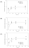
Effect of PCBs on the lactational transfer of methyl mercury in mice: PBPK modeling
- PMID: 20046988
- PMCID: PMC2707941
- DOI: 10.1016/j.etap.2008.08.014
Effect of PCBs on the lactational transfer of methyl mercury in mice: PBPK modeling
Abstract
MeHg and PCB exposure to lactating mice were analyzed and a physiologically-based pharmacokinetic (PBPK) model was developed to describe the lactational transfer of MeHg in mice. The influence of albumin on the lactational transfer of MeHg was incorporated into the PBPK model. Experimental results with lactating mice and their pups showed that co-exposure with PCB congeners increased the lactational transfer of MeHg to the pups, which was associated with the rise of albumin levels in maternal blood. Observed results were matched with PBPK model simulations conducted under the assumptions that (1) MeHg bound to plasma albumin is transferred to maternal milk, and (2) PCB congeners may increase the lactational transfer of MeHg by escalating albumin levels in maternal blood. Further refinement of PBPK model quantitatively described the pharmacokinetic changes of MeHg by co-exposure with PCBs in pup's tissues.
Keywords: MeHg; PBPK; PCBs; lactational transfer.
Conflict of interest statement
Conflict of Interest
Authors declare there is no conflict of interest with regards to the research described in the manuscript.
Figures


Similar articles
-
Comparison of pharmacokinetic interactions and physiologically based pharmacokinetic modeling of PCB 153 and PCB 126 in nonpregnant mice, lactating mice, and suckling pups.Toxicol Sci. 2002 Jan;65(1):26-34. doi: 10.1093/toxsci/65.1.26. Toxicol Sci. 2002. PMID: 11752682
-
A physiologically based pharmacokinetic model for lactational transfer of PCB 153 with or without PCB 126 in mice.Arch Toxicol. 2007 Feb;81(2):101-11. doi: 10.1007/s00204-006-0130-0. Epub 2006 Jul 21. Arch Toxicol. 2007. PMID: 16858609
-
Computer simulation of the lactational transfer of tetrachloroethylene in rats using a physiologically based model.Toxicol Appl Pharmacol. 1994 Apr;125(2):228-36. doi: 10.1006/taap.1994.1068. Toxicol Appl Pharmacol. 1994. PMID: 8171430
-
Using structural information to create physiologically based pharmacokinetic models for all polychlorinated biphenyls.Toxicol Appl Pharmacol. 1997 Jun;144(2):340-7. doi: 10.1006/taap.1997.8139. Toxicol Appl Pharmacol. 1997. PMID: 9194418 Review.
-
Effects of polychlorinated biphenyls on the nervous system.Toxicol Ind Health. 2000 Sep;16(7-8):305-33. doi: 10.1177/074823370001600708. Toxicol Ind Health. 2000. PMID: 11693948 Review.
Cited by
-
Modeling bioavailability to organs protected by biological barriers.In Silico Pharmacol. 2013 May 31;1:8. doi: 10.1186/2193-9616-1-8. eCollection 2013. In Silico Pharmacol. 2013. PMID: 25505653 Free PMC article. Review.
-
A generalized physiologically-based toxicokinetic modeling system for chemical mixtures containing metals.Theor Biol Med Model. 2010 Jun 2;7:17. doi: 10.1186/1742-4682-7-17. Theor Biol Med Model. 2010. PMID: 20525215 Free PMC article.
References
-
- Bemis JC, Seegal RF. Polychlorinated biphenyls and methylmercury alter intracellular calcium concentrations in rat cerebellar granule cells. Neurotoxicology. 2000;21:1123–1134. - PubMed
-
- Boersma ER, Lanting CI. Environmental exposure to polychlorinated biphenyls (PCBs) and dioxins. Consequences for longterm neurological and cognitive development of the child lactation. Adv Exp Med Biol. 2000;478:271–287. - PubMed
-
- Borlak J, Dangers M, Thum T. Aroclor 1254 modulates gene expression of nuclear transcription factors: implications for albumin gene transcription and protein synthesis in rat hepatocyte cultures. Toxicol Appl Pharmacol. 2002;181:79–98. - PubMed
-
- Bowers WJ, Nakai JS, Chu I, Wade MG, Moir D, Yagminas A, Gill S, Pulido O, Meuller R. Early developmental neurotoxicity of a PCB/organochlorine mixture in rodents after gestational and lactational exposure. Toxicol Sci. 2004;77:51–62. - PubMed
Grants and funding
LinkOut - more resources
Full Text Sources

