. 2010 Apr;9(2):147-61.
doi: 10.1111/j.1474-9726.2009.00545.x. Epub 2009 Dec 28.

Endogenous glucocorticoids decrease skeletal angiogenesis, vascularity, hydration, and strength in aged mice

Affiliations

Endogenous glucocorticoids decrease skeletal angiogenesis, vascularity, hydration, and strength in aged mice

Robert S Weinstein et al. Aging Cell. 2010 Apr.

Abstract

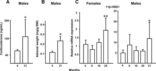
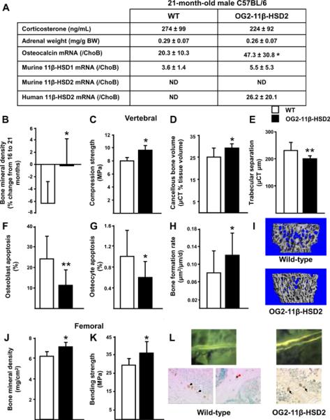
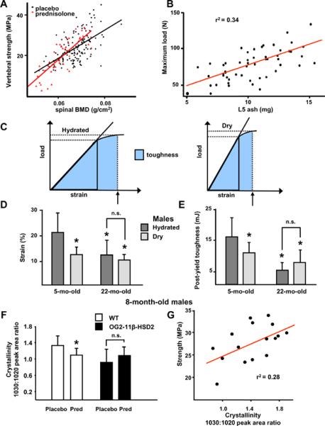
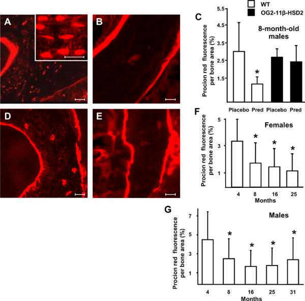
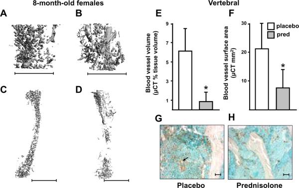
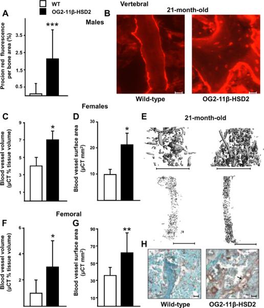
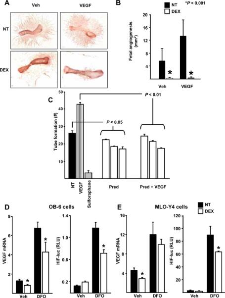
Aging or glucocorticoid excess decrease bone strength more than bone mass in humans and mice, but an explanation for this mismatch remains elusive. We report that aging in C57BL/6 mice was associated with an increase in adrenal production of glucocorticoids as well as bone expression of 11beta-hydroxysteroid dehydrogenase (11beta-HSD) type 1, the enzyme that activates glucocorticoids. Aging also decreased the volume of the bone vasculature and solute transport from the peripheral circulation to the lacunar-canalicular system. The same changes were reproduced by pharmacologic hyperglucocorticoidism. Furthermore, mice in which osteoblasts and osteocytes were shielded from glucocorticoids via cell-specific transgenic expression of 11beta-HSD type 2, the enzyme that inactivates glucocorticoids, were protected from the adverse effects of aging on osteoblast and osteocyte apoptosis, bone formation rate and microarchitecture, crystallinity, vasculature volume, interstitial fluid, and strength. In addition, glucocorticoids suppressed angiogenesis in fetal metatarsals and hypoxia inducible factor-1alpha transcription and vascular endothelial growth factor production in osteoblasts and osteocytes. These results, together with the evidence that dehydration of bone decreases strength, reveal that endogenous glucocorticoids increase skeletal fragility in old age as a result of cell autonomous effects on osteoblasts and osteocytes leading to interconnected decrements in bone angiogenesis, vasculature volume, and osteocyte-lacunar-canalicular fluid.

Keywords: 11β-hydroxysteroid dehydrogenase; Aging; angiogenesis; apoptosis; bone histomorphometry; glucocorticoids; hydraulic support; osteoporosis.


Figures

Figure 1
Figure 1
C57BL/6 mice from a cohort at Harlan Inc. maintained by NIA exhibit an increase in endogenous glucocorticoid production and activation with age. Increased serum corticosterone (n = 3 to 9 per group) (A) and adrenal weight (n = 10 to 12 per group) (B) from 4- to 31-month-old male mice are shown. Vertebral 11β-HSD1 expression in 4- to 25-month-old female mice (n = 10 to 12 per group) and in 4- to 31-month old male mice (n = 3 to 9 per group) (C) also increased with age. There was no significant change in ChoB with bone age. Data represent the mean ± SD. * P < 0.05 vs. 4-month-old mice, ** P < 0.005 vs. 4-month-old mice.
Figure 2
Figure 2
Preservation of bone density, strength, microarchitecture, osteoblast and osteocyte apoptosis, and formation rate in 21-month-old OG2-11β-HSD2 mice developed on a C57BL/6 background, bred, and maintained at UAMS (OG2-11β-HSD2, black bars) as compared to wild-type mice also maintained at UAMS (WT, white bars). Earlier development of endogenous hyperglucocorticoidism in the mice raised at UAMS as compared to those from the Harlan/NIA source is documented by the corticosterone levels, adrenal weights, and relative osteocalcin expression data. The expression of 11β-HSD1 in the wild-type and transgenic animals was similar. Murine 11β-HSD2 was not detectable in either wild-type or transgenic mice and human 11β-HSD2 was only detected in the transgenic animals as expected. (A), comparisons of the change in vertebral BMD from 16 to 21 months-of-age (B), compression strength (C), μCT determinations of bone volume/ tissue volume, trabecular separation, and representative μCT images of bone (D, E, I), osteoblast and osteocyte apoptosis (F, G), bone formation rate (H), femoral BMD at 21 months-of-age (J), femoral bending strength (K), and representative photomicrographs of bone formation and apoptosis (L) are shown. Black arrows indicate the brown, ISEL-positive, apoptotic osteoblasts located at the cancellous bone perimeter and red arrows indicate apoptotic osteocytes are buried in lacunae within mineralized bone. Data represent the mean ± SD. ND, not detectable, * P <0.05, ** P <0.02. (n = 6 to 11 per group).
Figure 3
Figure 3
Glucocorticoids decrease bone strength more than BMD. Normalized for bone size, vertebral compression strength (N/mm2 or MPa) and BMD (g/cm2) were measured in male and female Swiss Webster mice, aged 5 to 6 months and given placebo (black circles, n = 148) or prednisolone (red triangles, n = 47) for 28 days (A). The relationship between vertebral ash weight (mg) and maximal load (N) in 53 of the Swiss Webster placebo control mice from (A) is shown in (B). The relationships between the measurements of load, strain and toughness (the area under the load/strain curve) before and after vacuum drying are shown in (C). (D, E) Water contributes to fracture resistance in young but not old C57BL/6 male mice as measured by bone strain or deformation (D) and bone toughness or resistance to fracture (E). Dark gray bars are hydrated and light gray bars are vacuum dried vertebrae. (n = 12 to 15 per group; * P < 0.05 vs. the hydrated 5-month-old group; n.s., not significant). (F, G) Fourier transform infrared imaging analysis in 8-month-old male wild-type (white bars) and OG2-11β-HSD2 transgenic mice (black bars) given placebo or prednisolone for 28 days. Crystallinity, the ratio of the relative areas of the 1030 and 1020 cm-1 peaks, decreased in wild-type but not in OG2-11β-HSD2 mice (* P < 0.05, n = 8 to 9 per group) (F). Vertebral compression strength and crystallinity were directly related (G). (r2 = 28%, r = 0.53, P < 0.03, n = 4 from each group). Data are given as the mean ± SD.
Figure 4
Figure 4
Prednisolone administration or aging decrease solute transport through the vertebral lacunarcanalicular system. Procion red fluorescence per cancellous bone area decreased after 28 days of prednisolone administration to 8-month-old male wild-type (WT, white bars) but this effect was blocked in the OG2-11β-HSD2 mice (black bars) (A-C). Eight-month-old WT and OG2-11β-HSD2 mice receiving placebo had similar procion fluorescence. Scale bars: 10 um. Procion red fluorescence decreased from 4- to 25-months-of-age in female C57BL/6 mice (D-F) and in 4- to 31-month-old males (G). Representative photomicrographs are shown. Data represent the mean ± SD. (* P < 0.05, n = 3 per group).
Figure 5
Figure 5
Bone vasculature decreases with prednisolone administration. Representative images of vertebrae and femora after perfusion with lead chromate administered to 8-month-old female C57BL/6 mice after 28 days of placebo (A and C) or prednisolone (B and D) administration (n = 5 per group). Scale bars: 1 mm. Comparisons of the vertebral blood vessel volume (E) and surface area (F) in mice receiving placebo (white bars) or prednisolone (pred, grey bars) are shown. * P < 0.05. Data indicate the mean ± SD. Representative photomicrographs of immunostaining for von Willebrand factor in sections of cancellous vertebral bone taken from the 8-month-old C57BL/6 mice after placebo (G) or prednisolone (H). Prednisolone reduced the vascularity by an average of 74.5%. Scale bars: 10 um. The arrow in (G) points to ring-shapes typical of cross-sections of blood vessels.
Figure 6
Figure 6
Preservation of bone lacunar-canalicular fluid, vascular volume, and vascular surface area in 21-month-old OG2-11β-HSD2 (black bars) compared to wild-type mice (white bars). Comparison of solute transport through the vertebral lacunar-canalicular system of male animals is shown in (A) and representative photomicrographs are shown in (B). Scale bars: 10 um. Comparisons of vertebral (C, D) and femoral (F, G) blood vessels after perfusion with lead chromate and representative μCT images from female animals (E). Scale bars: 1 mm. Data represent the mean ± SD. * P <0.05, ** P <0.02, *** P <0.002. (n = 6 to 11 per group. For measurements of procion red fluorescence and vertebral vessels, n = 3 per group). Representative photomicrographs of immunostaining for von Willebrand factor in sections of cancellous vertebral bone are shown in (H). The 21-month-old transgenics had an average of 77.8% greater vascular staining as compared with the similarly aged wild-type mice. Scale bars: 10 um. The arrow points to a longitudinal section of blood vessel.
Figure 7
Figure 7
Glucocorticoids inhibit angiogenesis and interfere with VEGF message and action. Fetal metatarsal endothelial sprouting with or without added VEGF was inhibited by 10–8 M dexamethasone (DEX) in wild-type animals (A, B). (n = 10 to 12 per group). Prednisolone decreases tube-like structure formation by cultured human umbilical vein endothelial cells (C). (NT) indicates no treatment, (VEGF) is vascular endothelial growth factor, (PRED) is 10 to 1000 nM of prednisolone, and sulforaphane is a negative control that suppresses endothelial cell proliferation. In OB-6 osteoblastic cells, 10–8 M DEX decreased basal as well as desferrioxamine (DFO)-stimulated VEGF mRNA levels and decreased DFO-stimulated HIF1α expression (D). In MLO-Y4 osteocytic cells, 10–8 M DEX also decreased basal VEGF mRNA levels and DFO-stimulated HIF1α expression (E). Data represent the mean ± SD. * P < 0.05.

Similar articles

Cited by

References

    1. Almeida M, Han L, Martin-Millan M, Plotkin LI, Stewart SA, Roberson PK, Kousteni S, O'Brien CA, Bellido T, Parfitt AM, Weinstein RS, Jilka RL, Manolagas SC. Skeletal involution by age-associated oxidative stress and its acceleration by loss of sex steroids. J. Biol. Chem. 2007a;282:27285–27297. - PMC - PubMed
    1. Almeida M, Han L, Martin-Millan M, O'Brien CA, Manolagas SC. Oxidative stress antagonizes Wnt signaling in osteoblast precursors by diverting beta-catenin from T cell factor- to forkhead box O-mediated transcription. J. Biol. Chem. 2007b;282:27298–27305. - PubMed
    1. Almeida M, Ambrogini E, Han L, Manolagas SC, Jilka RL. Increased lipid oxidation causes oxidative stress, increased PPAR{gamma} expression and diminished proosteogenic Wnt signaling in the skeleton. J. Biol. Chem. 2009 doi: 10.1074/jbc.M109.023572. - PMC - PubMed
    1. Ambrogini E, O'Brien CA, Martin-Millan M, Paik J, DePinho RA, Han L, Warren A, Shelton RS, Qui X, Goellner JJ, Jilka RL, Almeida M, Manolagas SC. Loss or Gain of FoxO Function in Osteoclasts and Osteoblasts Alters the Rate of Apoptosis and BMD in Mice. J. Bone. Min. Res. 2008;23:S70.
    1. Chen W-T, Shih TT, Chen R-C, Lo S-Y, Chou C-T, Lee J-M, Tu H-Y. Vertebral bone marrow perfusion evaluated with dynamic contrast-enhanced MR imaging: significance of aging and sex. Radiology. 2001;220:213–218. - PubMed

Publication types

Substances