. 2010 Jan 4;11(1):R1.
doi: 10.1186/gb-2010-11-1-r1.

Genomic characterization of the Yersinia genus

Affiliations

Genomic characterization of the Yersinia genus

Peter E Chen et al. Genome Biol. .

Abstract

Background: New DNA sequencing technologies have enabled detailed comparative genomic analyses of entire genera of bacterial pathogens. Prior to this study, three species of the enterobacterial genus Yersinia that cause invasive human diseases (Yersinia pestis, Yersinia pseudotuberculosis, and Yersinia enterocolitica) had been sequenced. However, there were no genomic data on the Yersinia species with more limited virulence potential, frequently found in soil and water environments.

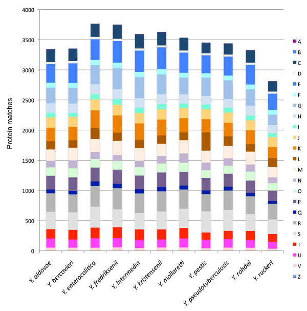
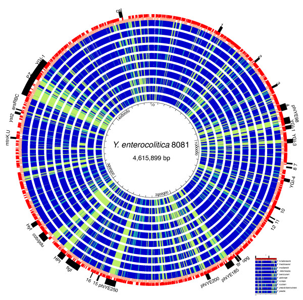
Results: We used high-throughput sequencing-by-synthesis instruments to obtain 25- to 42-fold average redundancy, whole-genome shotgun data from the type strains of eight species: Y. aldovae, Y. bercovieri, Y. frederiksenii, Y. kristensenii, Y. intermedia, Y. mollaretii, Y. rohdei, and Y. ruckeri. The deepest branching species in the genus, Y. ruckeri, causative agent of red mouth disease in fish, has the smallest genome (3.7 Mb), although it shares the same core set of approximately 2,500 genes as the other members of the species, whose genomes range in size from 4.3 to 4.8 Mb. Yersinia genomes had a similar global partition of protein functions, as measured by the distribution of Cluster of Orthologous Groups families. Genome to genome variation in islands with genes encoding functions such as ureases, hydrogenases and B-12 cofactor metabolite reactions may reflect adaptations to colonizing specific host habitats.

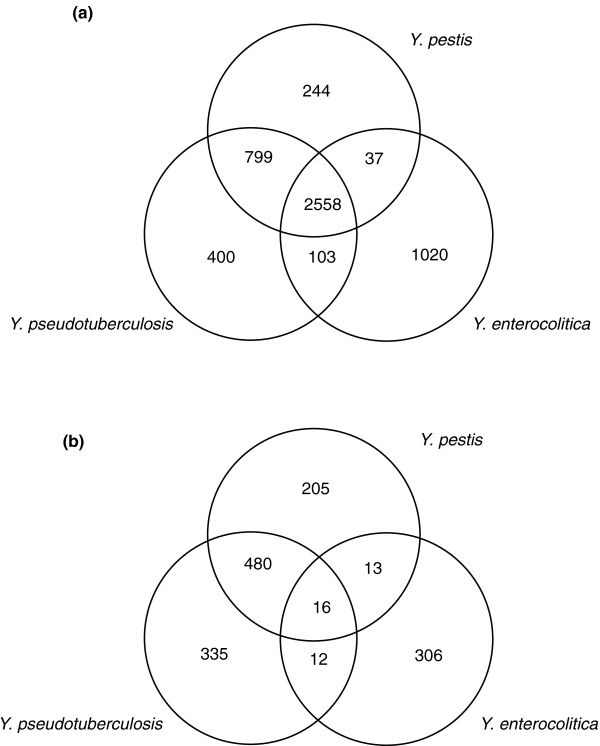
Conclusions: Rapid high-quality draft sequencing was used successfully to compare pathogenic and non-pathogenic members of the Yersinia genus. This work underscores the importance of the acquisition of horizontally transferred genes in the evolution of Y. pestis and points to virulence determinants that have been gained and lost on multiple occasions in the history of the genus.


Figures

Figure 1
Figure 1
Yersinia whole-genome phylogeny. The phylogeny of the Yersinia genus was constructed from a dataset of 681 concatenated, conserved protein sequences using the Neighbor-Joining (NJ) algorithm implemented by PHYLIP [51]. The tree was rooted using E. coli. The scale measures number of substitutions per residue. Tree topologies computed using maximum likelihood and parsimony estimates are identical with each other and the NJ tree (Additional file 20). The only branches not supported in more than 99% of the 1,000 bootstrap replicates using both methods are marked with asterisks. Both these branches were supported by >57% of replicates.
Figure 2
Figure 2
Comparison of major COG groups in Yersinia genomes. Bars represent the number of proteins assigned to COG superfamilies [52] for each genome, based on matches to the Conserved Domain Database [95] database with an E-value threshold <10-10. The COG groups are: U, intracellular trafficking; G, carbohydrate transport and metabolism; R, general function prediction; I, lipid transport and metabolism; D, cell cycle control; H, coenzyme transport and metabolism; B, chromatin structure; P, inorganic ion transport and metabolism; W, extracellular structures; O, post-translational modification; J, translation; A, RNA processing and editing; L, replication, recombination and repair; C, energy production; M, cell wall/membrane biogenesis; Q, secondary metabolite biosynthesis; Z, cytoskeleton; V, defense mechanisms; E, amino acid transport and metabolism; K, transcription; N, cell motility; T, signal transduction; F, nucleotide transport; S, function unknown.
Figure 3
Figure 3
Distribution of protein clusters across Y. enterocolitica 8081, Y. pestis CO92, and Y. pseudotuberculosis IP32953. (a) The Venn diagram shows the number of protein clusters unique or shared between the two other high virulence Yersinia species (see Materials and methods). (b) The number of shared and unique clusters that do not contain a single member of the eight low human virulence genomes sequenced in this study.
Figure 4
Figure 4
Protein-based comparison of Y. enterocolitica 8081 to the Yersinia genus. The map represents the blast score ratio (BSR) [98,99] to the protein encoded by Y. enterocolitica [15]. Blue indicates a BSR >0.70 (strong match); cyan 0.69 to 0.4 (intermediate); green <0.4 (weak). Red and pink outer circles are locations of the Y. enterocolitica genes on the + and - strands. The genomes are ordered from outside to inside based on the greatest overall similarity to Y. enterocolitica: Y. kristensenii, Y. frederiksenii, Y. mollaretii, Y. intermedia, Y. bercovieri, Y. aldovae, Y. rohdei, Y. ruckeri, Y. pseudotuberculosis, and Y. pestis. The black bars on the outside refer to genome islands in Y. enterocolitica identified by Thomson et al. [15].

Similar articles

Cited by

References

    1. Ecker DJ, Sampath R, Willett P, Wyatt JR, Samant V, Massire C, Hall TA, Hari K, McNeil JA, Buchen-Osmond C, Budowle B. The Microbial Rosetta Stone Database: a compilation of global and emerging infectious microorganisms and bioterrorist threat agents. BMC Microbiol. 2005;5:19. doi: 10.1186/1471-2180-5-19. - DOI - PMC - PubMed
    1. Achtman M, Zurth K, Morelli G, Torrea G, Guiyoule A, Carniel E. Yersinia pestis, the cause of plague, is a recently emerged clone of Yersinia pseudotuberculosis. Proc Natl Acad Sci USA. 1999;96:14043–14048. doi: 10.1073/pnas.96.24.14043. - DOI - PMC - PubMed
    1. van Baarlen P, van Belkum A, Summerbell RC, Crous PW, Thomma BP. Molecular mechanisms of pathogenicity: how do pathogenic microorganisms develop cross-kingdom host jumps? FEMS Microbiol Rev. 2007;31:239–277. doi: 10.1111/j.1574-6976.2007.00065.x. - DOI - PubMed
    1. Van Ert MN, Easterday WR, Huynh LY, Okinaka RT, Hugh-Jones ME, Ravel J, Zanecki SR, Pearson T, Simonson TS, U'Ren JM, Kachur SM, Leadem-Dougherty RR, Rhoton SD, Zinser G, Farlow J, Coker PR, Smith KL, Wang B, Kenefic LJ, Fraser-Liggett CM, Wagner DM, Keim P. Global Genetic Population Structure of Bacillus anthracis. PLoS ONE. 2007;2:e461. doi: 10.1371/journal.pone.0000461. - DOI - PMC - PubMed
    1. Zwick ME, McAfee F, Cutler DJ, Read TD, Ravel J, Bowman GR, Galloway DR, Mateczun A. Microarray-based resequencing of multiple Bacillus anthracis isolates. Genome Biol. 2005;6:R10. doi: 10.1186/gb-2004-6-1-r10. - DOI - PMC - PubMed

Publication types

LinkOut - more resources