

Review
doi: 10.1098/rsta.2009.0248.
Multi-scale heat and mass transfer modelling of cell and tissue cryopreservation
Affiliations
- PMID: 20047939
- PMCID: PMC3263795
- DOI: 10.1098/rsta.2009.0248
Item in Clipboard
Review
Multi-scale heat and mass transfer modelling of cell and tissue cryopreservation
Philos Trans A Math Phys Eng Sci.
.
Abstract
Cells and tissues undergo complex physical processes during cryopreservation. Understanding the underlying physical phenomena is critical to improve current cryopreservation methods and to develop new techniques. Here, we describe multi-scale approaches for modelling cell and tissue cryopreservation including heat transfer at macroscale level, crystallization, cell volume change and mass transport across cell membranes at microscale level. These multi-scale approaches allow us to study cell and tissue cryopreservation.
Figures
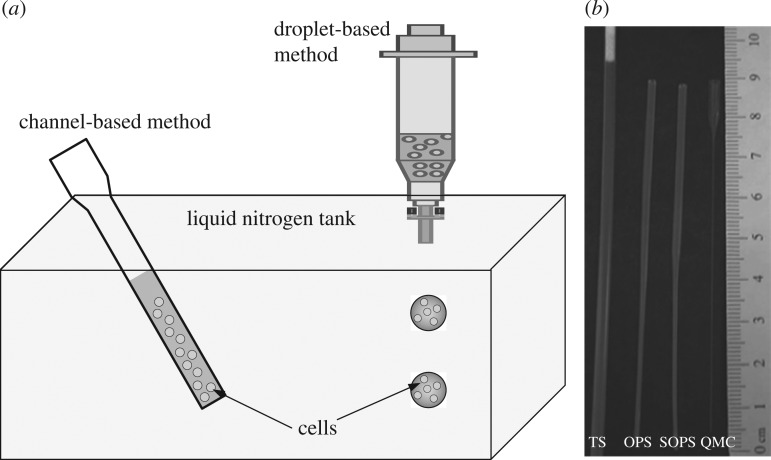
Channel-based and droplet-based methods for cryopreservation. (a) In channel-based methods, cells are mixed with media, e.g. CPAs, inside a channel. The whole channel is then immersed in liquid nitrogen for freezing. In droplet-based methods, cell-laden droplets are ejected into nitrogen for freezing (Demirci & Montesano 2007a). (b) A comparison of the devices used in channel-based methods (Arav et al. 2002; He et al. 2008): the traditional straw (TS), the open-pulled straw (OPS), the superfine open-pulled straw (SOPS) and the quartz micro-capillary (QMC). Adapted from He et al. (2008).
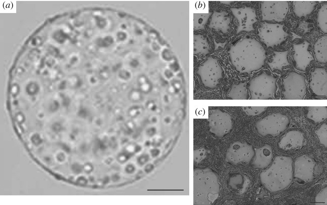
Droplet-based cryopreservation is a promising method as shown from in vivo results. (a) Post-thawed morphology of microencapsulated myoblasts in alginate gel droplet with 10% DMSO. Histological analysis (hematoxylin and eosin staining) 180 days post-implantation shows (c) the myoblasts cryopreserved in microcapsules explanted from the subcutaneous tissue of individuals compared with (b) non-cryopreserved control. Freezing protocol used: 1 h at −20 °C; 23 h at −80 °C; liquid nitrogen (−196 °C) for 44 days. Adapted from Murua et al. (2009). Scale bars: (a) 100 μm, (b,c) 200 μm.
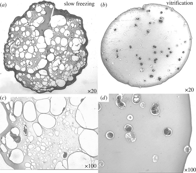
Morphology of frozen and vitrified pancreatic substitute beads at −90 °C. The beads comprise TC3 cells encapsulated in alginate gel. Beads frozen using a conventional controlled-rate (−1 °C min−1) protocol with 1 M DMSO show considerable ice formation throughout the construct (white spaces a,c). In contrast, beads vitrified with VS55 appear to be ice free (b,d). At higher magnification (c,d), the encapsulated individual cells and clusters of cells appear to be shrunken and compressed within the frozen matrix (c) compared with the more normal appearance of the cells encapsulated in the vitrified matrix (d). Adapted from Song et al. (2005).
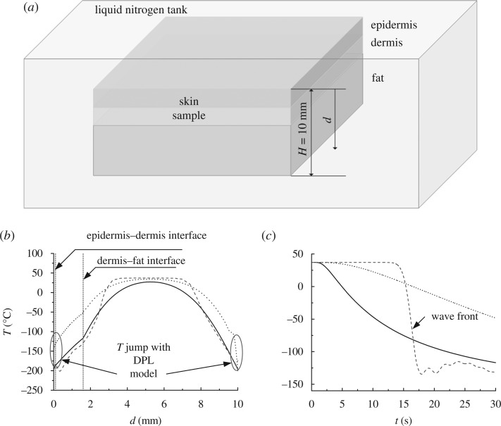
Modelling of tissue cryopreservation. Different models (i.e. Fourier model, thermal wave model and DPL model) are used to indicate the non-Fourier effect on heat transfer process during tissue cryopreservation. (a) Schematic of skin tissue immersed in liquid nitrogen for freezing. (b) Temperature distribution within skin at t=30 s. (c) Temperature variation with time at dermis–fat interface at r=1.6 mm. (b,c) Solid line, τq=0 s and τT=0 s; dashed line, τq=10 s and τT=0 s; and dotted line, τq=10 s and τT=10 s.
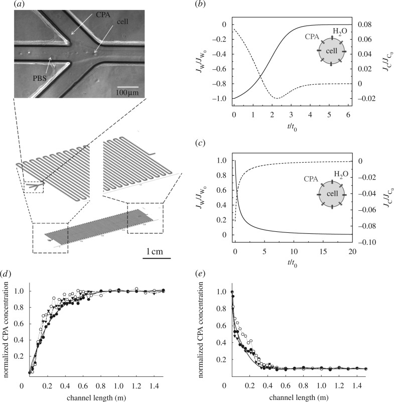
Modelling of CPA uploading and unloading processes using a microfluidic device for cell cryopreservation. (a) The microfluidic device has a three-input channel with dimensions of 100 μm in height, 100 μm in width and 1.5 m in length. Bright field image is the microfluidic channel inlet during flow. (b) Dimensionless water fluxes and CPA fluxes across cell membrane during the CPA loading step and (c) CPA unloading step. Here, V0 is the initial water volume in the cell, C0 is the final CPA concentration, t0=V0/(A0RTC0Lp) is the characteristic time, Jw0=V0/(A0t0) is the initial water flux and Jc0= C0/(A0t0) is the initial CPA flux. Normalized CPA concentration variation along the microfluidic channel in the (d) loading step and (e) unloading step agrees well with experimental data. (b,c) Solid line, water flux; dotted line, CPA flux. (d,e) Filled circle, experiment run 1; open circle, experiment run 2; inverted triangle, experiment run 3; solid line, numerical. Adapted from Song et al. (2009).
Similar articles
-
Thermal-mechanical deformation modelling of soft tissues for thermal ablation.Biomed Mater Eng. 2014;24(6):2299-310. doi: 10.3233/BME-141043. Biomed Mater Eng. 2014. PMID: 25226930
-
Lattice Boltzmann method for solving the bioheat equation.Phys Med Biol. 2008 Feb 7;53(3):N15-23. doi: 10.1088/0031-9155/53/3/N01. Epub 2008 Jan 14. Phys Med Biol. 2008. PMID: 18199898
-
Energy Dissipation in Ex Vivo Porcine Liver During Electrosurgery.IEEE Trans Biomed Eng. 2017 Jun;64(6):1211-1217. doi: 10.1109/TBME.2016.2595525. Epub 2016 Jul 27. IEEE Trans Biomed Eng. 2017. PMID: 27479955 Free PMC article.
-
Ratio of entropy to enthalpy in thermal transitions in biological tissues.J Biomed Opt. 2006 Jul-Aug;11(4):041108. doi: 10.1117/1.2343437. J Biomed Opt. 2006. PMID: 16965136 Review.
-
A review of heat transfer in human tooth--experimental characterization and mathematical modeling.Dent Mater. 2010 Jun;26(6):501-13. doi: 10.1016/j.dental.2010.02.009. Epub 2010 Mar 19. Dent Mater. 2010. PMID: 20303579 Review.
Cited by
-
Crystallisation Degree Analysis during Cryopreservation of Biological Tissue Applying Interval Arithmetic.Materials (Basel). 2023 Mar 9;16(6):2186. doi: 10.3390/ma16062186. Materials (Basel). 2023. PMID: 36984066 Free PMC article.
-
Engineering hydrogels as extracellular matrix mimics.Nanomedicine (Lond). 2010 Apr;5(3):469-84. doi: 10.2217/nnm.10.12. Nanomedicine (Lond). 2010. PMID: 20394538 Free PMC article. Review.
-
Testicular cryopreservation: From technical aspects to practical applications.Histol Histopathol. 2025 Jul;40(7):967-978. doi: 10.14670/HH-18-869. Epub 2025 Jan 2. Histol Histopathol. 2025. PMID: 39810720 Review.
-
Nanoliter droplet vitrification for oocyte cryopreservation.Nanomedicine (Lond). 2012 Apr;7(4):553-64. doi: 10.2217/nnm.11.145. Epub 2011 Dec 21. Nanomedicine (Lond). 2012. PMID: 22188180 Free PMC article.
-
Prediction and control of number of cells in microdroplets by stochastic modeling.Lab Chip. 2012 Nov 21;12(22):4884-93. doi: 10.1039/c2lc40523g. Lab Chip. 2012. PMID: 23034772 Free PMC article.
References
-
- Agca Y. 2000. Cryopreservation of oocyte and ovarian tissue. ILAR J 41, 207–220.. - PubMed
-
- Antaki P. J. 2005. New interpretation of non-Fourier heat conduction in processed meat. J. Heat. Transf. 127, 189–193.. (10.1115/1.1844540) - DOI
-
- Antinori M., Licata E., Dani G., Cerusico F., Versaci C., Antinori S. 2007. Cryotop vitrification of human oocytes results in high survival rate and healthy deliveries. Reprod. Biomed. Online 14, 72–79. - PubMed
-
- Arav A., Yavin S., Zeron Y., Natan D., Dekel I., Gacitua H. 2002. New trends in gamete’s cryopreservation. Mol. Cell. Endocrinol 187, 77–81.. - PubMed
Publication types
MeSH terms
Grants and funding
LinkOut - more resources
Full Text Sources
