. 2010 Apr;38(7):2399-410.
doi: 10.1093/nar/gkp1194. Epub 2010 Jan 4.

A novel mechanism for the scission of double-stranded DNA: BfiI cuts both 3'-5' and 5'-3' strands by rotating a single active site

Affiliations

A novel mechanism for the scission of double-stranded DNA: BfiI cuts both 3'-5' and 5'-3' strands by rotating a single active site

Giedrius Sasnauskas et al. Nucleic Acids Res. 2010 Apr.

Abstract

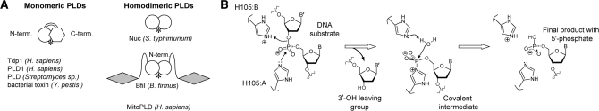
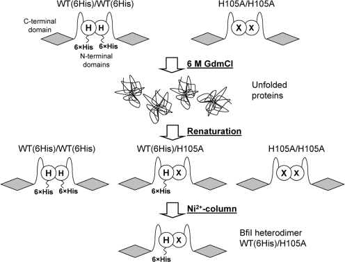
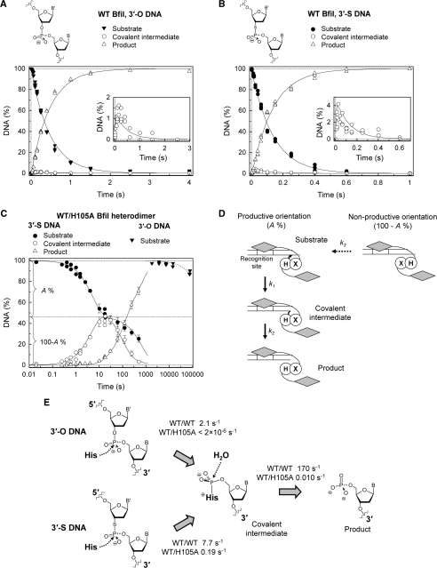
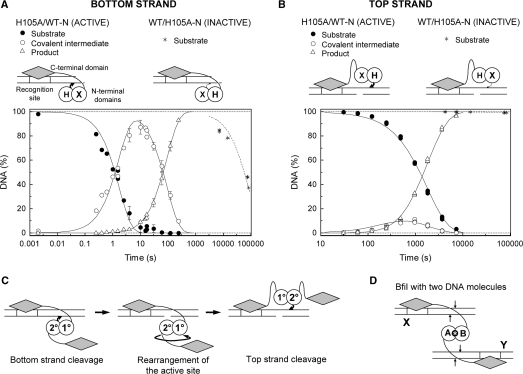
Metal-dependent nucleases that generate double-strand breaks in DNA often possess two symmetrically-equivalent subunits, arranged so that the active sites from each subunit act on opposite DNA strands. Restriction endonuclease BfiI belongs to the phospholipase D (PLD) superfamily and does not require metal ions for DNA cleavage. It exists as a dimer but has at its subunit interface a single active site that acts sequentially on both DNA strands. The active site contains two identical histidines related by 2-fold symmetry, one from each subunit. This symmetrical arrangement raises two questions: first, what is the role and the contribution to catalysis of each His residue; secondly, how does a nuclease with a single active site cut two DNA strands of opposite polarities to generate a double-strand break. In this study, the roles of active-site histidines in catalysis were dissected by analysing heterodimeric variants of BfiI lacking the histidine in one subunit. These variants revealed a novel mechanism for the scission of double-stranded DNA, one that requires a single active site to not only switch between strands but also to switch its orientation on the DNA.


Figures

Figure 1.
Figure 1.
Enzymes of the phospholipase D superfamily. (A). Monomeric and homodimeric PLD enzymes. The single PLD domain in the dimeric enzymes is depicted as a white circle, and the two domains in the monomeric enzymes as a circle and a hexagon. The DNA recognition domains of BfiI are marked as shaded diamonds. Both types of enzymes contain a single active site at the domain or subunit interface (marked by an asterisk). (B) The putative reaction mechanism of BfiI. During the first step of the reaction, His105 from subunit A (H105:A) attacks the scissile phosphate to generate the covalent intermediate, while His105 from subunit B (H105:B) protonates the 3′-leaving group. During the second step, a water molecule resolves the covalent intermediate releasing the first histidine (H105:A); the second histidine (H105:B) may facilitate this reaction by subtracting a proton from the water molecule.
Figure 2.
Figure 2.
Generation of heterodimeric variants of BfiI. The dimeric forms of His-tagged WT BfiI and the H105A mutant are indicated. In both cases, the BfiI monomer is shown as two domains connected by a linker: a C-terminal DNA-binding domain (shaded diamond) and a N-terminal domain for dimerization and catalysis (unfilled circle); ‘H’ marks H105 in the WT dimer and ‘X’ marks the H105A substitution in the mutant. The His-tagged WT and the H105A homodimers are mixed and completely unfolded with 6 M GdmCl. Subsequent removal of the denaturant results in formation of three species: the two initial homodimers and the heterodimer. The heterodimer with a single His-tag is separated from the homodimeric forms of BfiI lacking the His-tag or bearing two His-tags by Ni2+-chelating chromatography.
Figure 3.
Figure 3.
Cleavage of truncated phosphodiester and 3′-phosphorothiolate DNA substrates by WT BfiI and the WT/H105A heterodimer. The reactions contained 50–100 nM enzyme dimer and 1–2 nM radiolabeled DNA. The samples were collected and processed as described in ‘Materials and Methods’ section. (A and B) WT enzyme on the phosphodiester (3′-O) and 3′-phosphorothiolate (3′-S) substrates, 14/15 and 14/15s respectively. Cartoons above the graphs depict scissile linkages of the corresponding substrates. The time courses of intact substrate (filled symbols), covalent intermediate (open circles) and final product (open triangles) are shown: multiple data points at each time indicate values from repeat experiments. The inserts magnify the covalent intermediate data (<2 and <4% of total DNA for 3′-O and 3′-S substrates, respectively). Solid lines in both panels are the best fit of the experimental data to Equation (1) using a shared value of k2 for both substrates. The optimal fit gave k1(3′-O) = 2.1 ± 0.1 s−1, k1(3′-S) = 7.7 ± 0.1 s−1 and k2 = 170 ± 30 s−1. (C and D) The BfiI WT/H105A heterodimer on the 14/15 and 14/15s substrates. The heterodimer cleaved the 3′-S substrate 14/15s (filled circles), to form the covalent intermediate (open circles) and the final product (open triangles). The cartoon depicts the putative course of this reaction: upon mixing heterodimeric enzyme with the 14/15s substrate (the white box in the duplex marks the recognition site), only a fraction (A%) of DNA is bound by the enzyme in its active orientation capable of catalysis; the remaining fraction of the substrate (100-A%) may be cleaved only after re-binding of the enzyme in the active orientation (a slow process described by the rate constant k3). Solid lines are the optimal fit of this scheme to the 14/15s DNA cleavage data. The determined parameters are: k1 = 0.19 ± 0.02 s−1, k2 = 0.010 ± 0.001 s−1, k3 = 0.0014 ± 0.0002 s−1 and A = 54 ± 3%. Reactions of the WT/H105A heterodimer on the 3′-O substrate 14/15 are also shown (filled triangles). A single exponential fit (dashed line) gave a rate constant of 2 × 10⁻⁶ s−1 for the decline in concentration of the 14/15 substrate. (E) The impact of the single H105A substitution on BfiI activity. Compared to WT BfiI (WT/WT), the WT/H105A heterodimer lacking one active-site histidine had virtually no activity on the phosphodiester substrate, but cleaved the 3′-phosphorothiolate substrate with a 40-fold decrease in the rate of covalent intermediate formation and a 17 000-fold decrease in the rate of covalent intermediate hydrolysis.
Figure 4.
Figure 4.
Orientation of the BfiI active site during cleavage of the bottom and the top DNA strands. Single turnover experiments were performed as in Figure 3 on oligonucleotide substrates bearing 3′-phosphorothiolate moieties at the scissile bond in either the bottom or the top strand of DNA duplex. Cartoons above the graphs depict the complexes formed by the truncated BfiI heterodimers with the corresponding duplexes. (A) Cleavage of the bottom strand of the 14/15s DNA by the BfiI heterodimers H105A/WT-N and WT/H105A-N. The heterodimer H105A/WT-N cleaved the substrate (filled circles) to the final product (open triangles) via the covalent enzyme–DNA intermediate (open circles). Solid lines are the best fit of the experimental data to Equation (1), which yielded k1 = 0.57 ± 0.07 s−1 and k2 = 0.011 ± 0.001 s−1. The preparation of WT/H105A-N caused only a residual level of DNA cleavage (asterisks). The best fit to a single exponential (dashed line) yielded a cleavage rate of 1.2 × 10⁻⁵ s−1. (B) Cleavage of the top strand of the 25s/NICK substrate. The H105A/WT-N heterodimer, that was active on the bottom strand (A), also cleaved the 25s/NICK DNA (filled circles) to the final product (open triangles) via the covalent intermediate (open circles). The fit to Equation (1) yielded k1 = 0.00056 ± 0.00001 s−1 and k2 = 0.0041 ± 0.0003 s−1. The WT/H105A-N heterodimer displayed only residual activity on this substrate (asterisks). The fit to an exponential (dashed line) yielded a cleavage rate of 1 × 10⁻⁷ s−1. (C) A model for the reactions of WT BfiI on the bottom and the top strand of a DNA duplex. The H105 residue from the same subunit of the homodimer, the 2° subunit not bound to the DNA makes the nucleophilic attacks on the target phosphodiester bonds in both bottom and top strands of the DNA. To match the anti-parallel orientation of the two strands, the N-terminal domains of BfiI must rotate by 180° between the two hydrolysis reactions. (D) Schematic representation of the synaptic complex of the BfiI endonuclease bound two copies of its recognition sequence. The two DNA duplexes are marked X and Y: boxed segments indicate recognition sites and the positions of the scissile phosphates marked by arrows. The two subunits of the dimeric protein are noted as A and B and the single active site is marked by an asterisk. All four of the target phosphodiester bonds are cleaved in this one active site in sequential reactions.

Similar articles

Cited by

References

    1. Horton NC. In: Protein-Nucleic Acid Interactions: Structural Biology. Rice PA, Correll CC, editors. Cambridge, UK: RSC Publishing; 2008.
    1. Beese LS, Steitz TA. Structural basis for the 3′-5′ exonuclease activity of Escherichia coli DNA polymerase I: a two metal ion mechanism. EMBO J. 1991;10:25–33. - PMC - PubMed
    1. Derbyshire V, Grindley ND, Joyce CM. The 3′-5′ exonuclease of DNA polymerase I of Escherichia coli: contribution of each amino acid at the active site to the reaction. EMBO J. 1991;10:17–24. - PMC - PubMed
    1. Connolly BA, Eckstein F, Pingoud A. The stereochemical course of the restriction endonuclease EcoRI-catalyzed reaction. J. Biol. Chem. 1984;259:10760–10763. - PubMed
    1. Elliott SL, Brazier J, Cosstick R, Connolly BA. Mechanism of the I DNA T:G-mismatch endonuclease (Vsr protein) probed with thiophosphate-containing oligodeoxynucleotides. J. Mol. Biol. 2005;353:692–703. - PubMed

Publication types

MeSH terms