. 2010 Mar 19;285(12):8621-7.
doi: 10.1074/jbc.M109.092353. Epub 2010 Jan 4.

Rheb GTPase controls apoptosis by regulating interaction of FKBP38 with Bcl-2 and Bcl-XL

Affiliations

Rheb GTPase controls apoptosis by regulating interaction of FKBP38 with Bcl-2 and Bcl-XL

Dongzhu Ma et al. J Biol Chem. .

Abstract

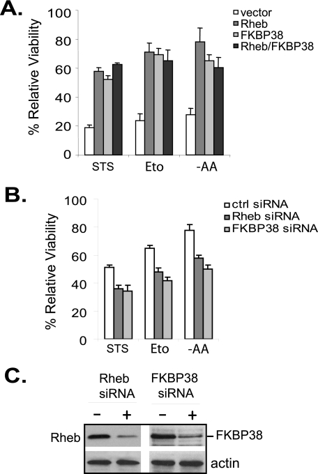
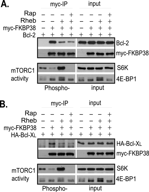
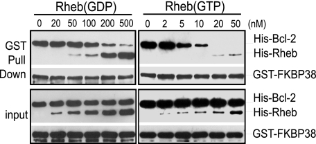
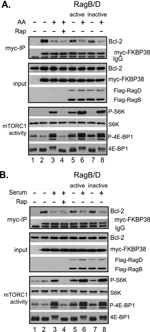
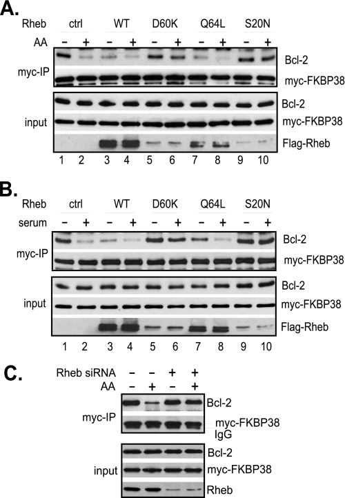
FKBP38 is a member of the family of FK506-binding proteins that acts as an inhibitor of the mammalian target of rapamycin (mTOR). The inhibitory action of FKBP38 is antagonized by Rheb, an oncogenic small GTPase, which interacts with FKBP38 and prevents its association with mTOR. In addition to the role in mTOR regulation, FKBP38 is also involved in binding and recruiting Bcl-2 and Bcl-X(L), two anti-apoptotic proteins, to mitochondria. In this study, we investigated the possibility that Rheb controls apoptosis by regulating the interaction of FKBP38 with Bcl-2 and Bcl-X(L). We demonstrate in vitro that the interaction of FKBP38 with Bcl-2 is regulated by Rheb in a GTP-dependent manner. In cultured cells, the interaction is controlled by Rheb in response to changes in amino acid and growth factor conditions. Importantly, we found that the Rheb-dependent release of Bcl-X(L) from FKBP38 facilitates the association of this anti-apoptotic protein with the pro-apoptotic protein Bak. Consequently, when Rheb activity increases, cells become more resistant to apoptotic inducers. Our findings reveal a novel mechanism through which growth factors and amino acids control apoptosis.


Figures

FIGURE 1.
FIGURE 1.
Overexpression of Rheb enhances cell survival under stress conditions. A, HeLa cells expressing an empty vector, FLAG-Rheb, Myc-FKBP38, or FLAG-Rheb and Myc-FKBP38 together were treated with 1 μm staurosporine (STS) for 6 h or 100 μm etoposide (Eto) for 20 h or starved for amino acids (−AA) for 16 h. Viability of the cells at the end of the treatment was assayed. B, HeLa cells were transfected with control (ctrl) siRNA or siRNAs specific for Rheb or FKBP38. Transfected cells were incubated for 72 before being treated with 500 nm staurosporine for 6 h or 100 μm etoposide for 16 h or starved for amino acids for 6 h. Data represent mean ± S.D. from three independent experiments. C, the levels of Rheb and FKBP38 in cells transfected with control siRNA (−) or Rheb-specific or FKBP38-specific siRNA (+) shown in B were determined by Western blotting using anti-Rheb or anti-FKBP38 antibody.
FIGURE 2.
FIGURE 2.
Rheb controls the interaction of FKBP38 with Bcl-2 and Bcl-XL. HeLa cells were transfected with the indicated genes. Transfected cells were treated with rapamycin (Rap+) or drug vehicle control (Rap−) for 30 min. Cells were lysed, and lysates were precipitated with anti-Myc antibody cross-linked to Protein A-agarose beads. The levels of Myc-FKBP38 and Bcl-2 (A) and hemagglutinin (HA)-Bcl-XL (B) in the precipitates (myc-IP) and lysates (input); the phosphorylation levels of S6 kinase (S6K) at Thr-389 and 4E-BP1 at Thr-37/46 (Phospho-); and their protein levels (input) in the lysates were determined by Western blotting.
FIGURE 3.
FIGURE 3.
Rheb regulates the interaction of FKBP38 with Bcl-2 in a GTP-dependent manner. Recombinant GST-FKBP38 was incubated with His-Bcl-2 in reactions containing the indicated concentrations of either GDP-loaded (left panels) or GTP-loaded (right panels) His-Rheb. The reaction mixtures were precipitated with glutathione-conjugated Sepharose beads. The amounts of GST-FKBP38, His-Bcl-2, and His-Rheb in the precipitates (GST Pull Down) and their levels in the reaction mixtures (input) were determined by Western blotting using anti-GST and anti-His antibodies.
FIGURE 4.
FIGURE 4.
Interaction of FKBP38 with Bcl-2 is regulated by amino acid and serum conditions. HeLa cells were transfected with vectors expressing S6 kinase, Bcl-2, and Myc-FKBP38, along with vector expressing FLAG-RagBGTP-RagDGDP (RagB/D active, lanes 5 and 6) or FLAG-RagBGDP-RagDGTP (RagB/D inactive, lanes 7 and 8) or empty vector (lanes 1–4). A, the transfected cells were starved for amino acids for 1 h followed by refeeding with amino acids (AA+) or vehicle control (AA−) for 30 min. B, the transfected cells were deprived of serum for 20 h followed by refeeding with serum (+) or vehicle control (−) for 30 min. Cells were treated with 40 nm rapamycin (Rap+) or drug vehicle (Rap−) for 30 min before refeeding. Cells were lysed at the end of the treatments, and lysates were precipitated with anti-Myc antibody (myc-IP) cross-linked with Protein A-agarose beads. The levels of Myc-FKBP38 and Bcl-2 in the precipitates and those of FKBP38, Bcl-2, and the Rag GTPases in the lysates (input); the phosphorylation levels of S6 kinase at Thr-389 (P-S6K) and 4E-BP1 at Thr-37/46 (P-4E-BP1); and their protein levels in the lysates were determined by Western blotting.
FIGURE 5.
FIGURE 5.
Rheb mediates the effect of amino acids and growth factors on the interaction of FKBP38 with Bcl-2. HeLa cells were transfected with Bcl-2 and Myc-FKBP38 together with wild-type FLAG-Rheb or its mutants. Transfected cells were either starved for amino acids for 1 h followed by amino acid repletion for 30 min (A) or deprived of serum for 20 h followed by readdition of serum for 30 min (B). C, HeLa cells were transfected with Rheb-specific siRNA or scrambled control siRNA. The transfected cells were starved for amino acids for 1 h followed by refeeding with amino acids (AA+) or vehicle control (AA−) for 30 min. Cells were lysed, and lysates were precipitated with anti-Myc antibody cross-linked with Protein A-agarose beads. The amounts of Myc-FKBP38 and Bcl-2 in the precipitates (myc-IP) and lysates (input) were determined by Western blotting. ctrl, control; WT, wild type.
FIGURE 6.
FIGURE 6.
Effect of Rheb and amino acids on the association of Bcl-XL with Bak. A, HeLa cells were starved for amino acids (AA) for 3 h and re-fed for 30 min. At 30 min prior to readdition of amino acids, cells were treated with 40 nm rapamycin (Rap) or drug vehicle control (ctrl). Cells were lysed, and mitochondrion-enriched fractions were obtained by fractionation. Upon solubilizing with detergent, the mitochondrial fraction was precipitated with anti-Bcl-XL antibody. The amounts of Bcl-XL and Bak in the precipitates (Bcl-XL-IP) and lysates (input) were determined by Western blotting. B, HeLa cells expressing control vector, FLAGwild-type (WT) FLAG-Rheb, or Rheb mutants were starved for amino acids for 3 h followed by refeeding with amino acids for 30 min. Cells were then collected and analyzed as in A. C, HeLa cells expressing control vector or FLAG-Rheb were exposed to 100 μm etoposide (+) or drug vehicle control (−) for 20 h followed by treatment with 40 nm rapamycin (+) or vehicle control (−) for 30 min. Cells were lysed and analyzed as in A.

Similar articles

Cited by

References

    1. Crino P. B., Nathanson K. L., Henske E. P. (2006) N. Engl. J. Med. 355, 1345–1356 - PubMed
    1. Mak B. C., Yeung R. S. (2004) Cancer Invest. 22, 588–603 - PubMed
    1. Li Y., Corradetti M. N., Inoki K., Guan K. L. (2004) Trends Biochem. Sci. 29, 32–38 - PubMed
    1. Manning B. D., Cantley L. C. (2003) Trends Biochem. Sci. 28, 573–576 - PubMed
    1. Inoki K., Corradetti M. N., Guan K. L. (2005) Nat. Genet. 37, 19–24 - PubMed

Publication types

MeSH terms