. 2010 Apr 16;285(16):12169-80.
doi: 10.1074/jbc.M109.087197. Epub 2010 Jan 4.

Identification of a key residue mediating bone morphogenetic protein (BMP)-6 resistance to noggin inhibition allows for engineered BMPs with superior agonist activity

Affiliations

Identification of a key residue mediating bone morphogenetic protein (BMP)-6 resistance to noggin inhibition allows for engineered BMPs with superior agonist activity

Kening Song et al. J Biol Chem. .

Abstract

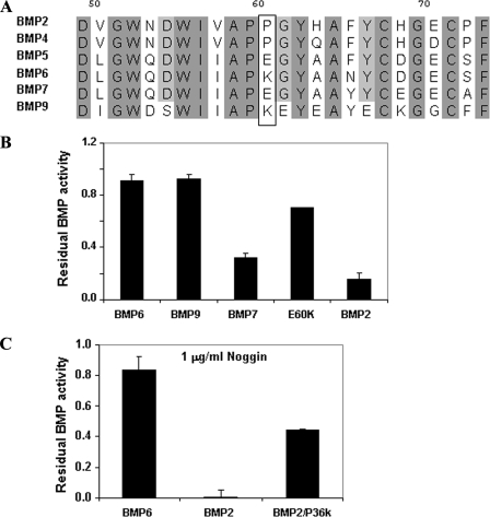
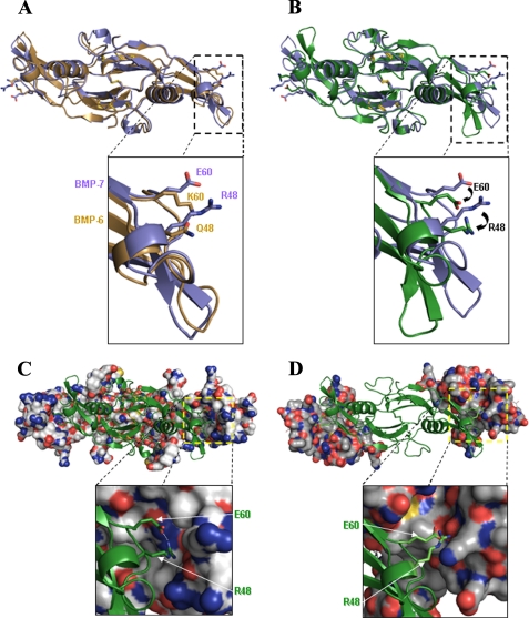
Bone morphogenetic proteins (BMPs) are used clinically to induce new bone formation in spinal fusions and long bone non-union fractures. However, large amounts of BMPs are needed to achieve these effects. BMPs were found to increase the expression of antagonists, which potentially limit their therapeutic efficacy. However, the relative susceptibility of osteoinductive BMPs to different antagonists is not well characterized. Here we show that BMP-6 is more resistant to noggin inhibition and more potent in promoting osteoblast differentiation in vitro and inducing bone regeneration in vivo when compared with its closely related BMP-7 paralog. Noggin was found to play a critical role as a negative feedback regulator of BMP-7 but not BMP-6-induced biological responses. Using BMP-6/7 chimeras, we identified lysine 60 as a key residue conferring noggin resistance within the BMP-6 protein. A remarkable correlation was found between the presence of a lysine at this position and noggin resistance among a panel of osteoinductive BMPs. Introduction of a lysine residue at the corresponding positions of BMP-2 and BMP-7 allowed for molecular engineering of recombinant BMPs with increased resistance to noggin antagonism.


Figures

FIGURE 1.
FIGURE 1.
Differential potency of BMP-6 and BMP-7 is determined by their difference in sensitivity to noggin inhibition. A, a panel of BMPs (BMP-2, BMP-4, BMP-5, BMP-6, BMP-7, GDF-5/BMP-14, and GDF-6/BMP-13) were tested for their ability to induce ALP activity in the rat osteosarcoma cell line, ROS 17/2.8. Each growth factor response was examined in a nine-point dose response in triplicates. Potency was assessed based on the EC50 derived from the non-linear regression of the mean result. B, BMP-6 is more potent than BMP-7 in stimulating BRE-transcriptional activity and alkaline phosphatase activity. Serum-starved C2C12 cells were transfected with BRE-luc reporter and stimulated with 25 ng/ml BMP-6, 25 ng/ml BMP-7, or vehicle control, and luciferase activity was analyzed 24 h post-transfection. Right panel, serum-starved C2C12 cells were stimulated with 100 ng/ml BMP-6, 100 ng/ml BMP-7, or vehicle control, and alkaline phosphatase activity was analyzed 48 h post-transfection. C, BMP-6 is more potent than BMP-7 in ectopic bone formation. Shown is an x-ray of critical size defects of rabbit ulnae treated with a collagen sponge (Helistat) alone (control; top), with rhBMP-6 (100 μg; middle), and with rhBMP-7 (100 μg; bottom) in vivo for 2 and 6 weeks after surgery. Micro-CT (μCT) analyses of critical size defects were performed in vivo at 8 weeks after surgery (cross-sections were horizontal and vertical). A, p < 0.05 versus C; B, p < 0.05 versus A (analysis of variance; Dunnett's test). RLU, relative luciferase units.
FIGURE 2.
FIGURE 2.
BMP-7 is more potent in inducing noggin mRNA expression than BMP-6. A and B, serum-starved C2C12 cells were stimulated with BMP-6 (50 ng/ml) or BMP-7 (50 ng/ml) or vehicle control for 18 h. Total RNA was extracted, and cDNA was prepared and expression of noggin was determined using quantitative PCR with expression being normalized to glyceraldehyde-3-phosphate dehydrogenase and expressed as -fold change relative to control. Non-stimulated controls are indicated with gray bars for BMP-6 and hatched bars for BMP-7. C, BMP-6-induced Smad1 phosphorylation is more sustained than BMP-7-induced Smad1 phosphorylation. Serum-starved C2C12 cells were stimulated with BMP-6 (50 ng/ml) or BMP-7 (50 ng/ml) for the indicated times, and cell lysates were analyzed by immunoblotting with phospho-Smad1/5 and total Smad1 antibodies. Upon noggin knockdown, BMP-7-induced responses are comparable with BMP-6-induced levels of Smad1 phosphorylation (D), BRE-luc activity (E), and ALP activity (F). C2C12 cells transfected with control siRNA or noggin siRNA were stimulated with BMP-6, BMP-7, or vehicle control. For Western blotting, cell lysates were analyzed by immunoblotting with phosphorylated Smad1/5 or total Smad1 antibody (E). For BRE-luc response, C2C12 cells were transfected with BRE-luc and analyzed for luciferase activity (E). For ALP activity, cell extracts were analyzed (F). Non-stimulated controls are indicated with gray bars for BMP-6 and hatched bars for BMP-7. RLU, relative luciferase units.
FIGURE 3.
FIGURE 3.
BMP-6 induction of downstream genes in primary human bone marrow-derived mesenchymal stem cells (hMSCs) is less susceptible to noggin inhibition than BMP-7. A, in order to compare BMP susceptibility to noggin inhibition, BMP-6, BMP-7, BMP-2, and BMP-4 were tested by an ALP assay in the ROS cells, in the presence of increasing concentrations of noggin. Each condition was tested in triplicates, and noggin dose-response curves were derived by fitting the data to a non-linear regression. The IC50 for noggin corresponding to each BMP was then calculated. These results indicate that the four BMPs tested are affected differently by noggin. BMP-4 is the most susceptible, followed by BMP-2, BMP-7, and then BMP-6. Although BMP-4, BMP-2, and BMP-7 are clearly sensitive to noggin inhibition, BMP-6 demonstrated marked resistance to noggin inhibition, with an IC50 value 4–16 times higher than that of other BMPs tested. B, BMP-6, but not BMP-7, is resistant to noggin-mediated inhibition. C2C12 cells transfected with BRE-luc reporter were challenged with BMP-6 or BMP-7 in the absence or presence of the indicated amounts of noggin and analyzed for luciferase activity. C, human mesenchymal stem cells were stimulated with BMP-6 or BMP-7, and effects on target genes, including Id-1, Dlx-5, Msx-2, and noggin, were measured by quantitative PCR. Induction of these target genes by BMP-7 was significantly inhibited by noggin in all cases. In particular, noggin inhibits its own induction by BMP-7 to a greater degree than other genes. In contrast, noggin did not affect induction of Id-1, Dlx-5, and noggin transcription induced by BMP-6. Noggin only marginally decreased the induction of Msx-2 by BMP-6. RLU, relative luciferase units.
FIGURE 4.
FIGURE 4.
Affinity measurements of noggin for BMPs using surface plasmon resonance. The affinity of recombinant noggin to BMPs was determined using surface plasmon resonance (BIAcore). Noggin-Fc was immobilized onto the sensor surface as described under “Experimental Procedures.” Free BMP-2, BMP-4, BMP-6, or BMP-7 was then applied at the indicated concentrations, at 75μl/min for 180 seconds followed by a 300-second dissociation phase. Blank buffer was used as a negative control. The dissociation constant Kd and the association constant Ka for each BMP were determined. The affinity to noggin-FC (KD) for each BMP was then calculated (listed in the table). RU, response units.
FIGURE 5.
FIGURE 5.
Noggin inhibits BMP-7 but not BMP-6 binding to BMP receptors. The indicated BMP receptors were transfected into COS cells and affinity-labeled by cross-linking with radiolabeled BMP-6 or BMP-7 in the absence or presence of noggin. Affinity-labeled BMP-BMP receptors were immunoprecipitated (IP) from cell extracts with ALK2- or ALK6-specific antibodies. Samples were washed, boiled in SDS sample buffer, and subjected to SDS-PAGE. Gels were dried and scanned with the STORM imaging system (Amersham Biosciences). BMPR-II, BMPR-II-BMP cross-linked complex; ALK2 and ALK6, ALK2-BMP and ALK6-BMP cross-linked complexes, respectively.
FIGURE 6.
FIGURE 6.
Identification of region in BMP-6 that is responsible for increased resistance to noggin inhibition. A, alignment of BMP-6 and BMP-7 mature domains. Shared amino acid residues are shaded. B, schematic representation of BMP-6 (shaded), BMP-7 (black), and BMP-6/7 chimeras. C, the chimera 80-1 is as resistant to noggin inhibition as BMP-6. D, the sensitivity of purified recombinant 80-1 chimera to noggin inhibition is comparable with that of BMP-6. 80-1 chimera was purified from the conditioned medium of transfected HEK 293 cells, and BMP-7 and BMP-6 were purified from stable Chinese hamster ovary expressing cell lines. A549-BRE cells were challenged with either BMP-6, BMP-7, or 80-1 proteins in the presence of increasing concentrations of noggin. Luciferase activity was measured 24 h post-BMP treatment, and the percentage inhibition was calculated for each noggin concentration.
FIGURE 7.
FIGURE 7.
Identification of a single residue in BMP-6 that is responsible for increased resistance to noggin inhibition. A, amino acid substitutions (at the positions indicated at the top) in the 80-1 chimera, in the various revertants (Rev) and in the BMP-7 mutants (E60K, Y65N, Y78H, and R48Q/E60K/Y65N). The corresponding amino acids in BMP-6 and BMP-7 are listed at the top. Amino acid substitutions are highlighted in boldface type. B, reverting Lys60 to Glu in the 80-1 chimera significantly reduces its sensitivity to noggin (1 μg/ml)-mediated repression of BRE-luc activity in A549-BRE cells. C, a single BMP-7 mutation at position 60 (E60K) significantly increases the protein resistance to noggin (1 μg/ml) inhibition. HEK293T cells were transfected with plasmids expressing BMP-7, BMP-6, 80-1, and each of the 80-1 revertants (B) or BMP-7 mutants (C), and equal volumes of the conditioned medium were used to induce luciferase expression in A549-BRE cells in the presence or absence of noggin (1 μg/ml). Residual BMP activity was determined 24 h post-A549-BRE cell treatment.
FIGURE 8.
FIGURE 8.
Generation of BMPs with superior agonistic activity. A, amino acid sequence alignment of the region extending from residue 49 to 73 (BMP-7 numbering) in the mature regions of BMP-2, -4, -5, -6, -7, and -9. B, the presence of a lysine at position 60 in BMP-6, BMP-9, and BMP-7 E60K is associated with resistance to noggin inhibition. The activity of purified recombinant BMP-2, BMP-6, BMP-9, BMP-7, and BMP-7 E60K was assessed using the A549-BRE cells. BMP-7 E60K was purified from the conditioned medium following a large scale transfection of HEK 293 cells. BMP-7, BMP-2, and BMP-6 were expressed in Chinese hamster ovary cells. BMP-9 was purchased from R&D Systems. A549-BRE cells were challenged with the purified recombinant proteins (100 ng/ml), in the presence (1 μg/ml) or absence of noggin. Luciferase activity was measured 24 h post-BMP treatment, and the residual BMP activity was calculated. C, BMP-2 mutant P36K is significantly more sensitive to noggin (1 μg/ml) inhibition than the wild type BMP-2.
FIGURE 9.
FIGURE 9.
Structural representation of BMP-7 interaction with noggin and ActR-II. A, structural comparison between BMP-6 (brown) and BMP-7 (purple). Images were generated using the PyMOL Molecular Graphics System (2002) (DeLano Scientific, LLC, Palo Alto, CA). The inset shows an enlarged view of the location of Glu60, Arg48, and Gln48 amino acid residues. B, BMP-7 structure when bound to noggin (green) or in the free state (purple). The inset shows an enlarged view of the location of Glu60 and Arg48 amino acid residues. C, BMP-7-noggin complex. BMP-7 is displayed as a green secondary structure schematic diagram, and noggin is shown as a molecular surface (white, carbon; red, oxygen; blue, nitrogen; yellow, sulfur). The inset shows an enlarged view of the location of Glu60 and Arg48 amino acid residues (stick display). D, BMP-7-ActRII complex. BMP-7 is displayed as a green secondary structure schematic diagram, and ActRII is shown as a molecular surface (white, carbon; red, oxygen; blue, nitrogen; yellow, sulfur). The inset shows an enlarged view of the locations of Glu60 and Arg48 residues (stick display).

Similar articles

Cited by

References

    1. Urist M. R. (1965) Science 150, 893–899 - PubMed
    1. Wozney J. M., Rosen V., Celeste A. J., Mitsock L. M., Whitters M. J., Kriz R. W., Hewick R. M., Wang E. A. (1988) Science 242, 1528–1534 - PubMed
    1. Sampath T. K., Reddi A. H. (1981) Proc. Natl. Acad. Sci. U.S.A. 78, 7599–7603 - PMC - PubMed
    1. Reddi A. H., Reddi A. (2009) Cytokine Growth Factor Rev. 20, 341–342 - PubMed
    1. Miyazono K., Maeda S., Imamura T. (2005) Cytokine Growth Factor Rev. 16, 251–263 - PubMed

Publication types

MeSH terms

LinkOut - more resources