. 2010 Mar 5;285(10):7805-17.
doi: 10.1074/jbc.M109.091173. Epub 2010 Jan 4.

Site-specific phosphorylation of CXCR4 is dynamically regulated by multiple kinases and results in differential modulation of CXCR4 signaling

Affiliations

Site-specific phosphorylation of CXCR4 is dynamically regulated by multiple kinases and results in differential modulation of CXCR4 signaling

John M Busillo et al. J Biol Chem. .

Abstract

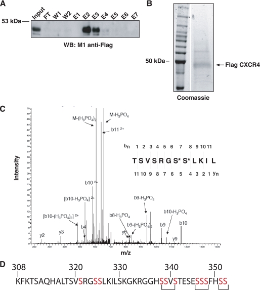
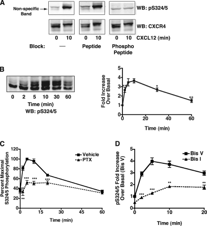
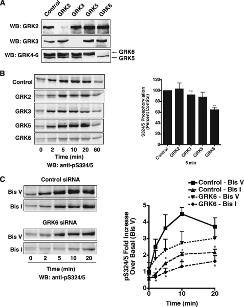
The chemokine receptor CXCR4 is a widely expressed G protein-coupled receptor that has been implicated in a number of diseases including human immunodeficiency virus, cancer, and WHIM syndrome, with the latter two involving dysregulation of CXCR4 signaling. To better understand the role of phosphorylation in regulating CXCR4 signaling, tandem mass spectrometry and phospho-specific antibodies were used to identify sites of agonist-promoted phosphorylation. These studies demonstrated that Ser-321, Ser-324, Ser-325, Ser-330, Ser-339, and two sites between Ser-346 and Ser-352 were phosphorylated in HEK293 cells. We show that Ser-324/5 was rapidly phosphorylated by protein kinase C and G protein-coupled receptor kinase 6 (GRK6) upon CXCL12 treatment, whereas Ser-339 was specifically and rapidly phosphorylated by GRK6. Ser-330 was also phosphorylated by GRK6, albeit with slower kinetics. Similar results were observed in human astroglia cells, where endogenous CXCR4 was rapidly phosphorylated on Ser-324/5 by protein kinase C after CXCL12 treatment, whereas Ser-330 was slowly phosphorylated. Analysis of CXCR4 signaling in HEK293 cells revealed that calcium mobilization was primarily negatively regulated by GRK2, GRK6, and arrestin3, whereas GRK3, GRK6, and arrestin2 played a primary role in positively regulating ERK1/2 activation. In contrast, GRK2 appeared to play a negative role in ERK1/2 activation. Finally, we show that arrestin association with CXCR4 is primarily driven by the phosphorylation of far C-terminal residues on the receptor. These studies reveal that site-specific phosphorylation of CXCR4 is dynamically regulated by multiple kinases resulting in both positive and negative modulation of CXCR4 signaling.


Figures

FIGURE 1.
FIGURE 1.
Establishing and characterizing HEK293 cells that stably express CXCR4. A, HEK293 cells or cells stably expressing FLAG CXCR4 were loaded with the ratiometric calcium indicator Fura-2/AM before stimulation with CXCL12 (100 nm). The change in intracellular calcium was calculated by monitoring the change in fluorescence of Fura-2/AM. Shown is a representative trace of calcium mobilization from three independent experiments. B, after a 6-h serum starvation, HEK293 cells or FLAG CXCR4 cells were stimulated with CXCL12 for the times indicated. pERK2 was normalized to total ERK2 by LiCor imaging to determine that overexpression of CXCR4 led to a 2.5-fold increase in ERK activation. Shown are representative Western blots (WB) of phospho-ERK1/2 and total ERK2 from three independent experiments. CXCL12-promoted retardation of electrophoretic mobility of endogenous CXCR4 (C) or FLAG tagged CXCR4 (D). After a 6-h serum starvation, cells were stimulated with 100 nm CXCL12 for the indicated times. Crude membranes were prepared, and 50 μg of solubilized protein (endogenous) or an equal volume of whole cell lysate (Flag CXCR4) was separated by 10% SDS-PAGE. Shown is a representative Western blot from four independent experiments.
FIGURE 2.
FIGURE 2.
Purification and mass spectrometry analysis of CXCR4. A, FLAG CXCR4 was purified from five 15-cm plates after a 10-min stimulation with 50 nm CXCL12. The bulk of the receptor (∼80%) eluted in fractions 2 and 3 and was highly purified as shown by Coomassie Blue staining (B). WB, Western blot. FT, flow through. C, shown is a representative mass spectrum of the peptide Thr-318 to Leu-328 after a chymotrypsin digest demonstrating that CXCR4 is phosphorylated on Ser-324 and Ser-325. D, shown is an amino acid sequence of the C-terminal tail of CXCR4. Residues highlighted in red are those that are predicted to be phosphorylated by mass spectrometry. Brackets under Ser-338-Ser-341, Ser-346-Ser-348, and Ser-351/Ser-352 indicate that one residue in each cluster is phosphorylated, although the exact residue was not identified by mass spectrometry.
FIGURE 3.
FIGURE 3.
Characterization of anti-Ser(P)-324/5 (pS324/5). A, shown is a representative Western blot (WB) demonstrating the specificity of the Ser(P)-324/5 antibody. Ten μg of purified antibody was incubated for 10 min with vehicle (PBS), 10 μg of peptide (C-Ahx-RGSSLKIL), or 10 μg of phosphopeptide (C-Ahx-RG(pS)(pS)LKIL) before overnight incubation with the nitrocellulose blots. B, cells stably expressing FLAG CXCR4 were stimulated at the time points indicated with 100 nm CXCL12. Lysates were processed and separated to visualize the agonist promoted gel shift of CXCR4. Blots were incubated overnight with a 1:1000 dilution of crude Ser(P)-324/5 antibody. Ser(P)-324/5 was normalized to total CXCR4, and data are presented as the -fold increase over basal (±S.E., n = 4). The -fold increase at 10 min is significantly different from 2, 30, and 60 min (p = 0.004, 0.05, and 0.009, respectively) but not 5 min. C, cells stably expressing FLAG CXCR4 were treated overnight with vehicle (PBS) or pertussis toxin (PTX, 100 ng/ml) before stimulation with 100 nm CXCL12. Ser(P)-324/5 was normalized to total CXCR4, and data are presented as the percent maximum of vehicle-treated cells (± S.E., n = 3). D, cells stably expressing FLAG CXCR4 were serum-starved for 6 h. 30 min before CXCL12 stimulation cells were pretreated with 2.5 μm Bis I or Bis V. Ser(P)-324/5 was normalized to total CXCR4, and data are presented as the -fold increase over basal (Bis V) (±S.E., n = 4; *, p ≤ 0.05; **, p ≤ 0.01; ***, p ≤ 0.001).
FIGURE 4.
FIGURE 4.
GRK6 contributes to Ser-324/5 phosphorylation after CXCL12 stimulation. A, shown is a representative Western blot (WB) demonstrating specific and efficient knockdown of GRKs endogenously expressed in HEK293 cells 72 h post-transfection. B, knockdown of GRK6, but not GRK2, GRK3, or GRK5 led to a significant reduction in phosphorylation of Ser-324/5. Left panel, shown is a representative Western blot using purified anti-Ser(P)-324/5. Right panel, shown is a comparison of Ser(P)-324/5 phosphorylation after a 5-min stimulation with CXCL12. Ser(P)-324/5 was normalized to total CXCR4, and data are presented as the percent of control at 5 min (±S.E., n = 4). C, GRK6 knockdown and PKC inhibition almost completely abolished phosphorylation of Ser-324/5. Cells transfected with GRK6 siRNA were pretreated with 2.5 μm Bis I or Bis V 30 min before stimulation with CXCL12. Left panel, shown is a representative Western blot using purified anti-Ser(P)-324/5. Right panel, Ser(P)-324/5 was normalized to total CXCR4, and data are presented as -fold increase over basal in control/Bis V-treated cells (±S.E., n = 3; *. p ≤ 0.05; **, p ≤ 0.01; ***, p ≤ 0.001).
FIGURE 5.
FIGURE 5.
Ser-330 and Ser-339 in CXCR4 are phosphorylated by GRK6. A, cells expressing FLAG CXCR4 were serum-starved for 6 h before stimulation with 100 nm CXCL12 for the times indicated. An equal volume of lysate was separated by SDS-PAGE and blotted with purified anti-Ser(P)-330 (top left panel) or anti-Ser(P)-339 (bottom left panel). Ser(P)-330 blots were processed to visualize the gel shift of CXCR4. Right panel, Ser(P)-330 or Ser(P)-339 blotting was normalized to total CXCR4, and data are presented as -fold increase over basal (±S.E., n = 4). For Ser-330, the -fold increase at 20 min was significantly different from 5, 10, and 60 min (p = 0.01, 0.02, and 0.004, respectively) but not 30 min. For Ser-339, the -fold increase at 2 min is significantly different from 5, 10, and 30 min (p = 0.01, 0.01, and 0.02, respectively). Seventy-two hours post-transfection, an equal volume of cell lysate was separated to visualize the gel shift of CXCR4 and blotted using purified anti-Ser(P)-330 (B) or anti-Ser(P)-339 (C). Shown are representative Western blots (WB) of four separate experiments.
FIGURE 6.
FIGURE 6.
GRKs differentially regulate signaling after activation of endogenous CXCR4 in HEK293 cells. A, HEK293 cells were loaded with the ratiometric calcium indicator Fura-2A/M 72 h after siRNA transfection. Cells were stimulated with 100 nm CXCL12, and changes in intracellular calcium were calculated from changes in fluorescence. Left panel, shown is a representative trace from six separate experiments. Right panel, shown is the mean ± S.E. increase in peak calcium transient calculated from six separate experiments. B, shown is the effect of GRK knockdown on CXCL12-mediated activation of ERK1/2. Seventy-two hours post-transfection cells were serum-starved for 6 h before stimulation with CXCL12 (100 nm). Shown is a representative Western blot (WB) from five independent experiments. C, left panel, pERK2 was normalized to total ERK2, and data are presented as the percent maximal ERK2 activation as compared with control (±S.E., n = 4). Right panel, comparison of maximal ERK2 activation (5 min) after stimulation with CXCL12 (100 nm) (±S.E., n = 4; *, p ≤ 0.05; **, p ≤ 0.01; ***, p ≤ 0.001).
FIGURE 7.
FIGURE 7.
Non-visual arrestins differentially regulate CXCR4-mediated signaling. A, mean (±S.E.) increase in peak calcium transient after stimulation of endogenous CXCR4 calculated from seven separate experiments is shown. B, mean (±S.E.) increase in peak calcium transient after stimulation of FLAG CXCR4 calculated from three separate experiments is shown. C and D, shown is the effect of arrestin knockdown on ERK1/2 activation after activation of HEK293 (C) or FLAG CXCR4 (D) cells. Seventy-two hours post-transfection, cells were serum-starved for 6 h before stimulation with CXCL12 (100 nm). Left panels, shown are representative Western blots from seven (C) and four (D) separate experiments. Right panels, pERK2 was normalized to total ERK2, and data are presented as the percent maximal ERK2 activation as compared with control (± S.E.; *, p ≤ 0.05; **, p ≤ 0.01; ***, p ≤ 0.001).
FIGURE 8.
FIGURE 8.
Phosphorylation of CXCR4 differentially regulates the recruitment and conformation of the non-visual arrestins. A BRET assay was performed to determine whether CXCR4 was able to recruit the non-visual arrestins after activation. HEK-293T cells were transiently transfected with wild type (WT) RLucII-tagged CXCR4 and either GFP-tagged arrestin2 or arrestin3. CXCL12 stimulation resulted in the recruitment of both arrestin2 (A) and arrestin3 (B). Interestingly, alanine substitution of the proposed GRK2/3 sites (5A Tail) eliminated arrestin2 recruitment (C) and significantly decreased arrestin3 recruitment (D). In contrast, alanine substitution of the identified GRK6 sites (GRK6SA) resulted in an increase in the BRET ratio observed, suggesting an altered conformation of the arrestins (C and D).
FIGURE 9.
FIGURE 9.
Regulation of CXCR4 activity and signaling. A, upon ligand binding, CXCR4 activates a number of signaling cascades resulting in calcium release from intracellular stores and phosphorylation of ERK1/2. B, phosphorylation of CXCR4 by GRK6 (Ser-324/5, Ser-330, and Ser-339) and GRK2 (residues between Ser-346 and Ser-352) resulted in the recruitment of arrestin3 to CXCR4, thereby attenuating G protein activation and calcium release. PLCβ, phospholipase Cβ; PIP2, phosphatidylinositol disphosphate; IP3, inositol trisphosphate. C, phosphorylation of CXCR4 by GRK6 (Ser-324/5, Ser-330, and Ser-339) and GRK3 (residues between Ser-346 and Ser-352) resulted in a conformation of arrestin2 that allows for full activation of ERK1/2. In contrast, GRK2 inhibits ERK1/2 activation most likely by regulating the activity of MEK (25).

Similar articles

Cited by

References

    1. Busillo J. M., Benovic J. L. (2007) Biochim. Biophys. Acta 1768, 952–963 - PMC - PubMed
    1. Hernandez P. A., Gorlin R. J., Lukens J. N., Taniuchi S., Bohinjec J., Francois F., Klotman M. E., Diaz G. A. (2003) Nat. Genet. 34, 70–74 - PubMed
    1. Feng Y., Broder C. C., Kennedy P. E., Berger E. A. (1996) Science 272, 872–877 - PubMed
    1. Zlotnik A. (2006) Contrib. Microbiol. 13, 191–199 - PubMed
    1. Diaz G. A., Gulino A. V. (2005) Curr. Allergy Asthma Rep. 5, 350–355 - PubMed

Publication types

MeSH terms