. 2010 Jan 19;121(2):293-305.
doi: 10.1161/CIRCULATIONAHA.109.871905. Epub 2010 Jan 4.

Intracoronary administration of cardiac progenitor cells alleviates left ventricular dysfunction in rats with a 30-day-old infarction

Affiliations

Intracoronary administration of cardiac progenitor cells alleviates left ventricular dysfunction in rats with a 30-day-old infarction

Xian-Liang Tang et al. Circulation. .

Abstract

Background: Administration of cardiac progenitor cells (CPCs) 4 hours after reperfusion ameliorates left ventricular function in rats with acute myocardial infarction (MI). Clinically, however, this approach is not feasible, because expansion of autologous CPCs after acute MI requires several weeks. Therefore, we sought to determine whether CPCs are beneficial in the more clinically relevant setting of an old MI (scar).

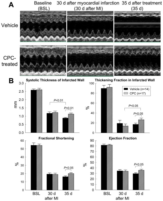
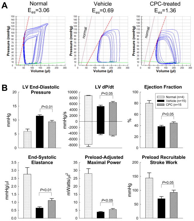
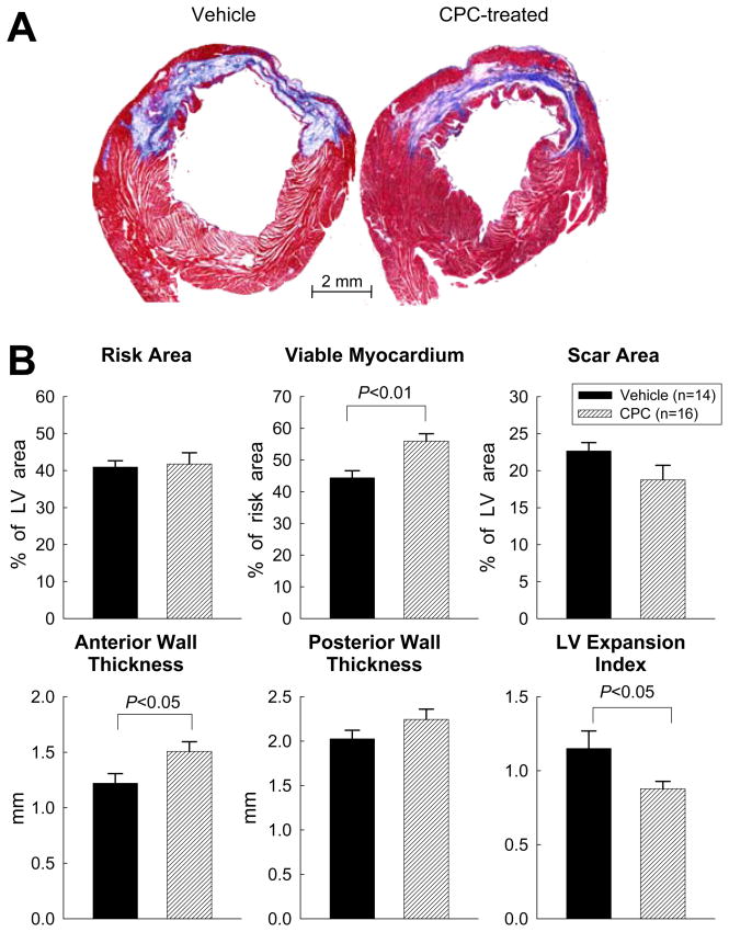
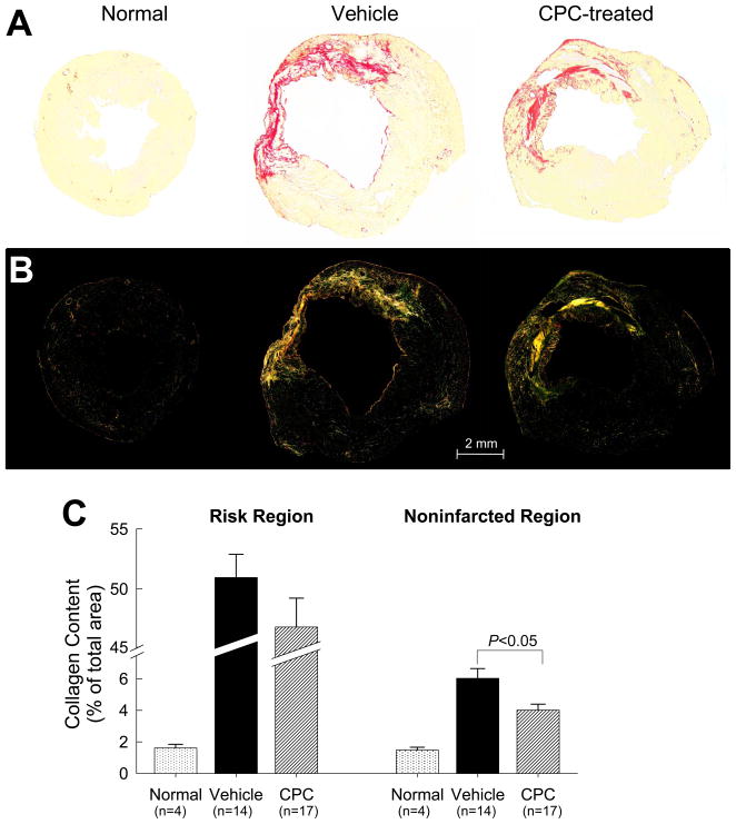
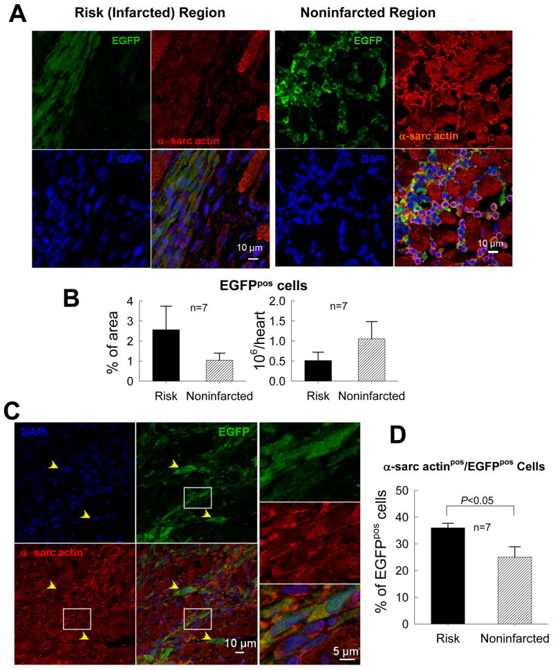
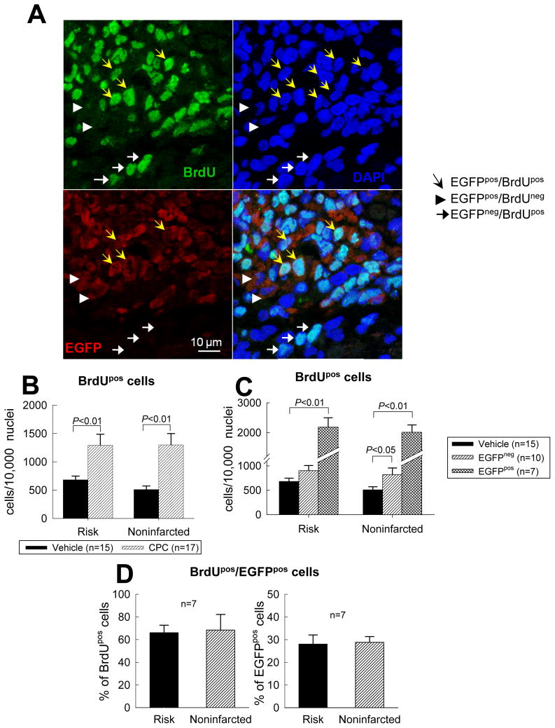
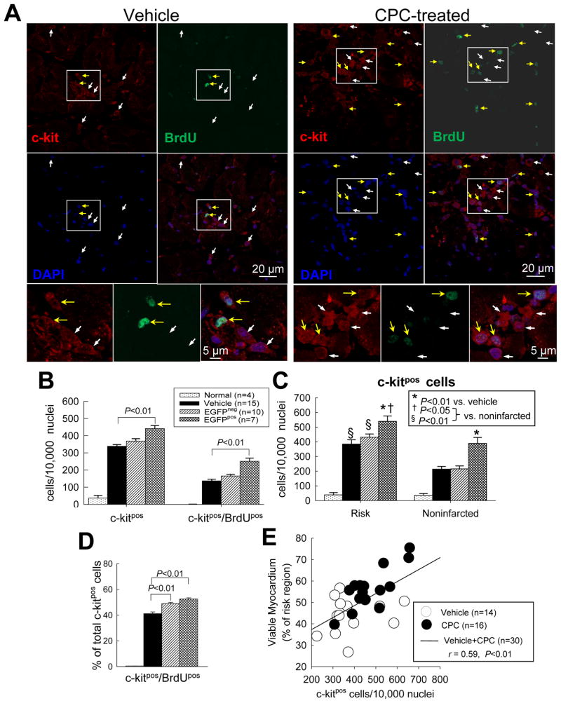
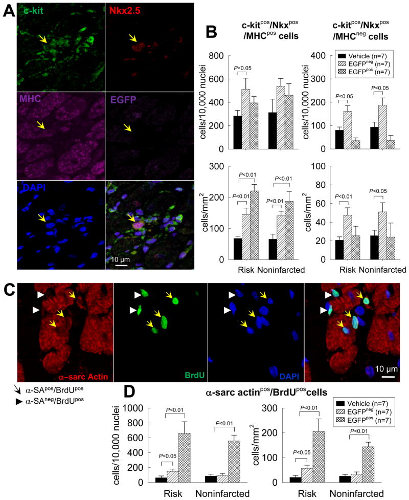
Methods and results: One month after coronary occlusion/reperfusion, rats received an intracoronary infusion of vehicle or enhanced green fluorescent protein-labeled CPCs. Thirty-five days later, CPC-treated rats exhibited more viable myocardium in the risk region, less fibrosis in the noninfarcted region, and improved left ventricular function. Cells that stained positive for enhanced green fluorescent protein that expressed cardiomyocyte, endothelial, and vascular smooth muscle cell markers were observed only in 7 of 17 treated rats and occupied only 2.6% and 1.1% of the risk and noninfarcted regions, respectively. Transplantation of CPCs was associated with increased proliferation and expression of cardiac proteins by endogenous CPCs.

Conclusions: Intracoronary administration of CPCs in the setting of an old MI produces beneficial structural and functional effects. Although exogenous CPCs can differentiate into new cardiac cells, this mechanism is not sufficient to explain the benefits, which suggests paracrine effects; among these, the present data identify activation of endogenous CPCs. This is the first report that CPCs are beneficial in the setting of an old MI when given by intracoronary infusion, the most widely applicable therapeutic approach in patients. Furthermore, this is the first evidence that exogenous CPC administration activates endogenous CPCs. These results open the door to new therapeutic applications for the use of autologous CPCs in patients with old MI and chronic ischemic cardiomyopathy.


Figures

Figure 1
Figure 1. Echocardiographic assessment of LV function
(A) Representative M-mode echocardiographic images from a vehicle-treated and a CPC-treated rat recorded at baseline (BSL), at 30 d after MI (before vehicle or CPC treatment) (MI), and at 35 d after treatment (35 d). (B) Quantitative echocardiographic parameters including systolic thickness of the anterior (infarcted) wall, thickening fraction in the anterior (infarcted) wall, LV fractional shortening, and LV ejection fraction. Data are means ± SEM.
Figure 2
Figure 2. Hemodynamic assessment of LV function at 35 d after treatment
(A) Representative pressure-volume loops from a normal, a vehicle-treated, and a CPC-treated rat recorded during preload manipulation by a brief period of inferior vena cava occlusion. (B) Quantitative analysis of hemodynamic variables including LV end-diastolic pressure, dP/dt, ejection fraction, end-systolic elastance (Ees), preload-adjusted maximal power, and preload-recruitable stroke work. Data are means ± SEM.
Figure 3
Figure 3. Morphometric analysis
(A) Representative Masson’s trichrome-stained myocardial sections from a vehicle-treated and a CPC-treated rat. Scar tissue and viable myocardium are identified in blue and red, respectively. (B) Quantitative analysis of LV morphometric parameters (for definition, see supplemental Fig. 11). Data are means ± SEM.
Figure 4
Figure 4. Myocardial collagen content
Representative microscopic images of a normal, a vehicle-treated, and a CPC-treated heart obtained using regular (A) and polarized (B) light. (C) Collagen content expressed as percent of total area in the scarred and noninfarcted region. LV sections were stained with picrosirius red and collagen content was quantitated under polarized light. Data are means ± SEM.
Figure 5
Figure 5. Myocardial content and differentiation of transplanted CPCs
(A) Representative confocal microscopic images from a CPC-treated rat showing presence of transplanted CPCs in the risk (infarcted) and noninfarcted regions, as evinced from immunoreactivity for EGFP (green). Some EGFPpos cells also express α-sarcomeric actin (red). (B) Quantitation of EGFPpos cells in the risk and noninfarcted region, expressed as percent EGFPpos myocardial area and as total calculated number of EGFPpos cells/heart. The number of EGFPpos cells/heart was estimated by multiplying the number of EGFPpos cells per unit area by the estimated number of EGFPpos cells present through the thickness of each slice in which EGFPpos cells were found (estimated 100 cells per 2-mm thickness of slice). (C) Representative confocal microscopic images showing colocalization of EGFP and α-sarcomeric actin in several cells in the border zone (the area in the white box is magnified in the three panels on the right). Yellow arrows indicate EGFPpos cells that do not expressα-sarcomeric actin. (D) Quantitative analysis of α-sarcomeric actinpos/EGFPpos cells. Data are means ± SEM.
Figure 6
Figure 6. Proliferation of transplanted CPCs
(A) Representative confocal microscopic images from a CPC-treated rat showing BrdU uptake in the infarcted region during the last 2 weeks of life. Yellow arrows indicate EGFPpos cells that are BrdU positive; white arrowheads EGFPpos cells that are BrdU negative; and white arrows EGFPneg cells that are BrdU positive. (B, C, and D) Quantitative analysis of BrdUpos cells in the risk and noninfarcted regions. Panel B shows the number of BrdUpos cells in vehicle-treated and CPC-treated groups; panel C shows the same data except that the CPC-treated group is subdivided into the subgroups that did or did not exhibit EGFPpos cells (in the seven hearts with EGFPpos cells, BrdUpos cells were counted in the entire region examined, both in the EGFPpos and in the EGFPneg areas). (D) Colocalization of BrdU and EGFP immunoreactivity in the seven CPC-treated hearts that exhibited EGFPpos cells. Data are means ± SEM.
Figure 7
Figure 7. Effect of CPC transplantation on c-kitpos cells
(A) Representative confocal microscopic images from a vehicle- treated and a CPC-treated rat showing c-kitpos cells (red) and BrdUpos cells (green) in the risk region. Yellow arrows indicate c-kitpos cells that are BrdU positive; white arrows c-kitneg cells that are BrdU positive; (B) Quantitative analysis of c-kitpos cells and double positive (c-kitpos/BrdUpos) cells in normal, vehicle-treated, and CPC-treated hearts (the last group is subdivided into two subgroups, that with EGFPpos cells [EGFPpos] and that without EGFPpos cells [EGFPneg]). (C) Quantitative analysis of c-kitpos cells in risk and noninfarcted regions in normal, vehicle-treated, and CPC-treated hearts [as in (B), the last group is subdivided into EGFPpos and EGFPneg subgroups]. (D) Quantitative analysis of double positive (c-kitpos/BrdUpos) cells expressed as a percent of all c-kitpos cells. (E) Correlation between the number of c-kitpos cells and the percent of viable myocardium in the risk region (r=0.59, P<0.01). Data are means ± SEM.
Figure 8
Figure 8. Cardiac commitment of endogenous CPCs after trasplantation of exogenous CPCs
The commitment of endogenous CPCs (c-kitpos/EGFPneg cells) to cardiac lineage was assessed by quantitating endogenous CPCs that expressed Nkx2.5 and MHC in vehicle- and CPC-treated hearts. New myocytes formed in the last 2 weeks of life were identified by measuring α-sarcomeric actinpos and BrdUpos cells. (A) Representative confocal microscopic image of a c-kitpos/EGFPneg/Nkx2.5pos/MHCpos cell obtained in serial sections of a CPC-treated heart. (B) Quantitative analysis of c-kitpos/EGFPneg/Nkx2.5pos/MHCpos cells in the risk and noninfarcted regions of vehicle- and CPC-treated hearts (the CPC-treated group is subdivided into two subgroups, one with EGFPpos cells [EGFPpos] and one without EGFPpos cells [EGFPneg]). The number of c-kitpos/EGFPneg/Nkx2.5pos/MHCpos cells is normalized to both number of cells (104 nuclei, upper panels) and area (mm2, lower panels). Because cell density in CPC-treated, EGFPpos hearts was higher (due to presence of clusters of EGFPpos cells), the magnitude of the increase in endogenous CPCs in CPC-treated, EGFPpos hearts was greater when the number of cells was normalized to area (mm2) than to number of nuclei. (C) Representative confocal microscopic image of α-sarcomeric actinpos/BrdUpos cells in a CPC-treated heart. (D) Quantitative analysis of the number of α-sarcomeric actinpos/BrdUpos cells in the risk and noninfarcted regions of vehicle- and CPC-treated hearts (subdivided into EGFPneg and EGFPpos subgroups). Data are means ± SEM.

References

    1. Dimmeler S, Zeiher AM, Schneider MD. Unchain my heart: the scientific foundations of cardiac repair. J Clin Invest. 2005;115:572–583. - PMC - PubMed
    1. Wollert KC, Drexler H. Clinical applications of stem cells for the heart. Circ Res. 2005;96:151–163. - PubMed
    1. Abdel-Latif A, Bolli R, Tleyjeh IM, Montori VM, Perin EC, Hornung CA, Zuba-Surma EK, Al-Mallah M, Dawn B. Adult bone marrow-derived cells for cardiac repair: a systematic review and meta-analysis. Arch Intern Med. 2007;167:989–997. - PubMed
    1. Bearzi C, Rota M, Hosoda T, Tillmanns J, Nascimbene A, De Angelis A, Yasuzawa-Amano S, Trofimova I, Siggins RW, Lecapitaine N, Cascapera S, Beltrami AP, D’Alessandro DA, Zias E, Quaini F, Urbanek K, Michler RE, Bolli R, Kajstura J, Leri A, Anversa P. Human cardiac stem cells. Proc Natl Acad Sci U S A. 2007;104:14068–14073. - PMC - PubMed
    1. Beltrami AP, Barlucchi L, Torella D, Baker M, Limana F, Chimenti S, Kasahara H, Rota M, Musso E, Urbanek K, Leri A, Kajstura J, Nadal-Ginard B, Anversa P. Adult cardiac stem cells are multipotent and support myocardial regeneration. Cell. 2003;114:763–776. - PubMed

Publication types

Substances