. 2010 Jan 18;207(1):77-84.
doi: 10.1084/jem.20091097. Epub 2010 Jan 4.

Heterozygous deficiency of delta-catenin impairs pathological angiogenesis

Affiliations

Heterozygous deficiency of delta-catenin impairs pathological angiogenesis

Laura M DeBusk et al. J Exp Med. .

Erratum in

  • J Exp Med. 2010 Feb 15;207(2):443
  • J Exp Med. 2010 Mar 15;207(3):669

Abstract

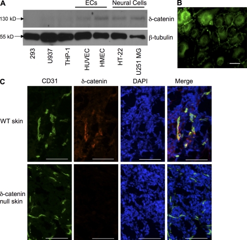
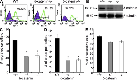
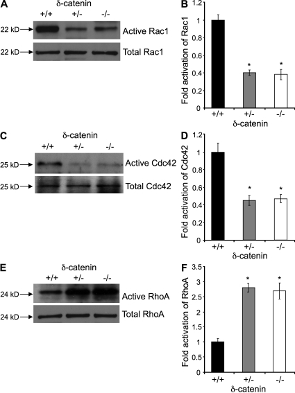
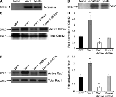
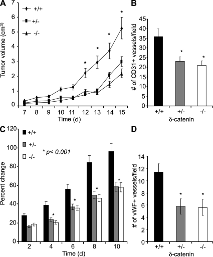
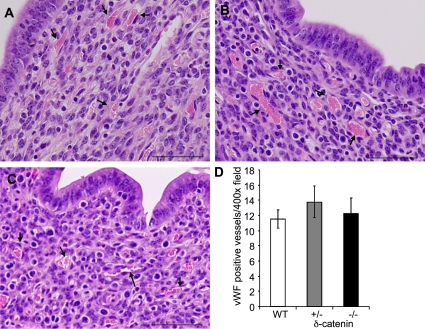
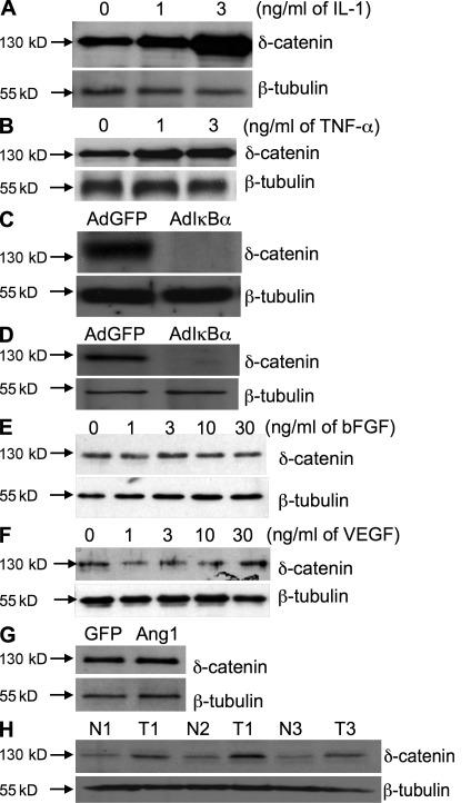
Vascular and neuronal networks share a similar branching morphology, and emerging evidence implicates common mechanisms in the formation of both systems. delta-Catenin is considered a neuronal catenin regulating neuron cell-cell adhesion and cell motility. Here, we report expression of delta-catenin in vascular endothelium, and show that deletion of only one allele of delta-catenin is sufficient to impair endothelial cell motility and vascular assembly in vitro and pathological angiogenesis in vivo, thereby inhibiting tumor growth and wound healing. In contrast, deletion of one or both allele of delta-catenin had no effects on hormone-induced physiological angiogenesis in the uterus. Molecular analysis confirmed a gene dosage effect of delta-catenin on Rho GTPase activity. Moreover, we show that inflammatory cytokines, but not angiogenic factors, regulate delta-catenin expression, and the levels of delta-catenin positively correlate to human lung cancers. Collectively, our data suggest that inflammation, commonly associated with disease conditions, induces delta-catenin expression that specifically regulates pathological, and not physiological, angiogenesis. Because only pathological angiogenesis is sensitive to decreased levels of delta-catenin, this may provide a good target for antiangiogenic therapy.


Figures

Figure 1.
Figure 1.
δ-Catenin is expressed in vascular endothelium. (A) Protein lysates collected from neuronal cells (HT-22 and U251 MG), endothelial cells (ECs; HUVEC and HMEC), epithelial cells (293), and leukocytes (THP-1 and U937) were subjected to Western blot analysis and probed with a δ-catenin–specific antibody. (B) β-tubulin was used as a loading control. Immunofluorescent staining for δ-catenin was performed in cultured HUVECs. (C) Mouse skin tissue sections from wild-type and δ-catenin–null mice were analyzed by immunofluorescent double staining using antibodies against CD31 for endothelium, and δ-catenin. Nuclei were stained with DAPI. 400× magnification. Each experiment was repeated three times, and representative images were shown. Bar, 200 µm.
Figure 2.
Figure 2.
δ-Catenin regulates endothelial cell motility and vascular tubule formation in vitro in a gene dosage–sensitive manner. (A) Microvascular endothelial cells were isolated and pooled from lungs (5 mice per group) of wild-type littermates, δ-catenin heterozygous-null, and homozygous-null mice. The endothelial cells were sorted with a CD31 antibody with a FACStarPlus flow cytometer. The percentage of CD31 positive cells was indicated in each graph. (B) The levels of δ-catenin in wild-type, δ-catenin+/−, and δ-catenin−/− endothelial cells were evaluated in cell lysates by Western blot. Endothelial cell migration and vascular tubule formation were measured in Transwell assay or Matrigel assay, respectively. (C) Migrated cells were counted after a 5-h incubation in 10 randomly selected high-power fields under microscopy. The data were collected from three independent experiments. Mean and SE were plotted. *, P < 0.01. (D) Vascular tubule formation was measured 18 h after cell plating. Vascular cross points were counted from 10 randomly selected high-power fields under microscopy. The data were collected from three independent experiments. *, P < 0.01. (E) Cell proliferation was measured by BrdU incorporation. The experiment was done in duplicate and repeated three times.
Figure 3.
Figure 3.
δ-Catenin regulates Rho GTPase activity in a gene dosage sensitive manner. Microvascular endothelial cells isolated from wild-type littermates, δ-catenin heterozygous-null, and homozygous-null mice were analyzed for Rho GTPase activity. Activation of Rac1 and Cdc42 was measured by PAK1-efficient pull-down assays. Precipitated activated Rac1 (A and B) or Cdc42 (C and D) and each total protein from cell lysate were analyzed by Western blot with specific antibody. RhoA activation was pulled down with Rhotekin-agarose beads, analyzed by Western blot, and probed with a RhoA-specific antibody (E and F). *, P < 0.001. Each experiment was repeated three times, and representative images were shown.
Figure 4.
Figure 4.
Vav1 interacts with δ-catenin and activates Cdc42 and Rac1. HUVECs were transfected with either an expression vector for control GFP or δ-catenin, or transfected with control shRNA or δ-catenin shRNA for 48 h. (A) Cell lysate was immunoprecipitated with antibody against Vav1 or control beads (none), and subjected to Western blot analysis with antibody against δ-catenin. Equal amount of cell lysate (lysate) was used as a loading control. (B) Cell lysate was also immunoprecipitated with antibody against δ-catenin or control beads (none) and subjected to Western blot analysis for Vav1. Equal amount of lysate (lysate) was used as a loading control. Cell lysates were analyzed for Cdc42 (C) and Rac1 (E) activation using PAK1-efficient pull-down assay. Precipitated and total protein was analyzed by Western blot. The levels of activated Cdc42 were compared with total levels of Cdc42 (D), and activated Rac1 to total levels of Rac1 (F). *, P < 0.05 and **, P < 0.01, compared with corresponding control. Each experiment was repeated three times, and representative images were shown.
Figure 5.
Figure 5.
Genetic deletion of δ-catenin in mice impairs pathological angiogenesis in a dose-dependent manner. 3LL tumor cells were injected subcutaneously into sex- and age-matched syngeneic δ-catenin homozygous-null, heterozygous-null, and wild-type littermates. (A) Tumor growth was measured by caliper for 15 d, and tumor volume was calculated and plotted (n = 10 mice/group; *, P < 0.001). (B) Tumor tissue sections from each group were analyzed by CD31 immunofluorescent staining, and CD31 positive vessels were counted from 10 randomly selected high-power fields under microscopy. Mean and SE were plotted. *, P < 0.05. (C) 6-mm biopsy punches were made in wild-type littermates, δ-catenin heterozygous-null, and δ-catenin homozygous-null mice. Wound size was measured daily. The percent changes of wound size over the original size were plotted (n = 7 mice/group; *, P < 0.01). (D) The vascular density measurement by vWF staining was performed in wounded tissues on day 3. Mean and SE were plotted. *, P < 0.01.
Figure 6.
Figure 6.
Deletion of δ-catenin in mice has no effect on hormone-induced angiogenesis in uterus. Female mice were superovulated, and the uteruses were harvested from wild-type littermates (A), heterozygous-null (B), and homozygous-null δ-catenin (C) mice. Hematoxylin and eosin staining was performed on tissue sections (A–C) for histological analysis. Arrows point to blood vessels. The tissue sections were also probed with a vWF antibody for vascular density. Positive vessels were counted from 10 randomly selected high-power fields under microscopy. Mean and SE were plotted (D). Bar, 200 µm.
Figure 7.
Figure 7.
Inflammatory cytokines induce the expression of δ-catenin through NF-κB in vascular endothelial cells. HUVECs were treated with increasing doses of either IL-1 (A) or TNF (B) for 24 h, and cell lysate was subjected to Western blotting for δ-catenin and β-tubulin expression. HUVECs were infected with adenoviral vectors expressing either GFP or a mutant IκBα for 24 h, followed by stimulation with recombinant IL-1 (C) or TNF (D) at 3 ng/ml for another 24 h. δ-Catenin expression was evaluated by Western blot. HUVECs were treated with increasing doses of either recombinant bFGF (E) or VEGF (F) for 24 h, and cell lysate was subjected to Western blotting for δ-catenin expression. (G) HUVECs were infected with adenoviral vector expressing GFP or Ang1 for 48 h, respectively. δ-Catenin expression was detected by Western blot. (H) Human lung tumor tissues (T) and adjacent normal lung tissues (N) from biopsies were subjected to Western blotting with δ-catenin or β-tubulin antibodies (n = 3 pairs).

References

    1. Bustelo X.R. 2000. Regulatory and signaling properties of the Vav family. Mol. Cell. Biol. 20:1461–1477 doi:10.1128/MCB.20.5.1461-1477.2000 - DOI - PMC - PubMed
    1. Carmeliet P., Tessier-Lavigne M. 2005. Common mechanisms of nerve and blood vessel wiring. Nature. 436:193–200 doi:10.1038/nature03875 - DOI - PubMed
    1. Eichmann A., Makinen T., Alitalo K. 2005. Neural guidance molecules regulate vascular remodeling and vessel navigation. Genes Dev. 19:1013–1021 doi:10.1101/gad.1305405 - DOI - PubMed
    1. Georgiades P., Ogilvy S., Duval H., Licence D.R., Charnock-Jones D.S., Smith S.K., Print C.G. 2002. VavCre transgenic mice: a tool for mutagenesis in hematopoietic and endothelial lineages. Genesis. 34:251–256 doi:10.1002/gene.10161 - DOI - PubMed
    1. Grosheva I., Shtutman M., Elbaum M., Bershadsky A.D. 2001. p120 catenin affects cell motility via modulation of activity of Rho-family GTPases: a link between cell-cell contact formation and regulation of cell locomotion. J. Cell Sci. 114:695–707 - PubMed

Publication types

MeSH terms