. 2010 Jun;51(6):1486-95.
doi: 10.1194/jlr.M003566. Epub 2010 Jan 4.

Strong induction of PCSK9 gene expression through HNF1alpha and SREBP2: mechanism for the resistance to LDL-cholesterol lowering effect of statins in dyslipidemic hamsters

Affiliations

Strong induction of PCSK9 gene expression through HNF1alpha and SREBP2: mechanism for the resistance to LDL-cholesterol lowering effect of statins in dyslipidemic hamsters

Bin Dong et al. J Lipid Res. 2010 Jun.

Abstract

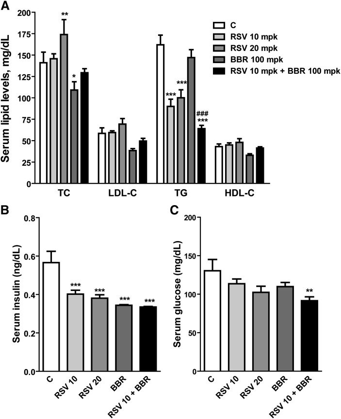
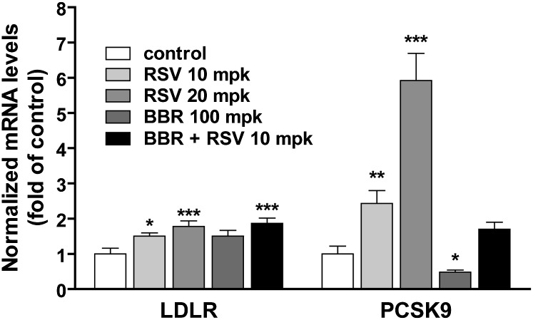
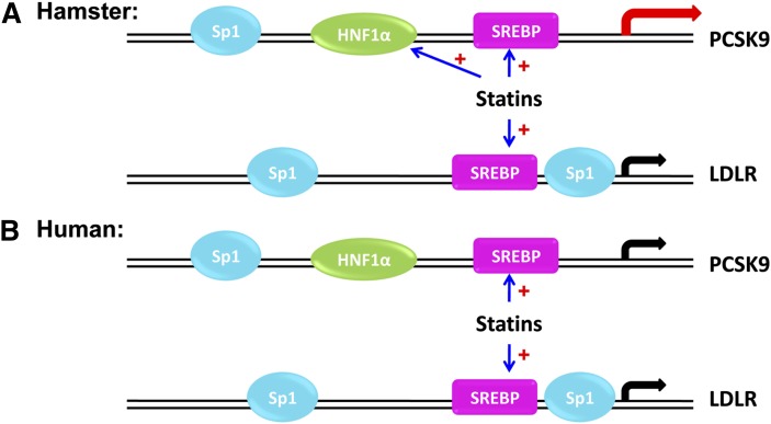
We investigated the role of proprotein convertase subtilisin/kexin type 9 (PCSK9) in the resistance of dyslipidemic hamsters to statin-induced LDL-cholesterol (LDL-C) reduction and the molecular mechanism by which statins modulated PCSK9 gene expression in vivo. We utilized the fructose diet-induced dyslipidemic hamsters as an in vivo model and rosuvastatin to examine its effects on liver PCSK9 and LDL receptor (LDLR) expression and serum lipid levels. We showed that rosuvastatin induced PCSK9 mRNA to a greater extent than LDLR mRNA in the hamster liver. The net result was that hepatic LDLR protein level was reduced. This correlated closely with an increase in serum LDL-C with statin treatment. More importantly, we demonstrated that in addition to an increase in sterol response element binding protein 2 (SREBP2) expression, rosuvastatin treatment increased the liver expression of hepatocyte nuclear factor 1 alpha (HNF1alpha), the newly identified key transactivator for PCSK9 gene expression. Our study suggests that the inducing effect of rosuvastatin on HNF1alpha is likely a underlying mechanism accounting for the higher induction of PCSK9 than LDLR because of the utilization of two transactivators (HNF1alpha and SREBP2) in PCSK9 transcription versus one (SREBP2) in LDLR transcription. Thus, the net balance is in favor of PCSK9-induced degradation of LDLR in the hamster liver, abrogating the effect of rosuvastatin on LDL-C lowering.


Figures

Fig. 1.
Fig. 1.
Differential modulation of lipid parameters by rosuvastatin and BBR. Hamsters on the fructose diet were administered vehicle (C), 10 mg/kg of rosuvastatin, 20 mg/kg of rosuvastatin, 100 mg/kg of BBR, and the combined treatment of rosuvastatin 10 mg/kg plus BBR at 100 mg/kg for 7 days. The serum samples were collected 24 h after the last treatment. TC, TG, LDL-C, HDL-C, and insulin were measured using commercial kits. Serum glucose was measured by a clinical chemistry analyzer (Olympus AU5431). Values are mean ± SEM of nine animals. *P < 0.05, ** P < 0.01, and *** P < 0.001 compared with the vehicle control group; ### P < 0.001 compared with rosuvastatin 10 mg/kg group (two-way ANOVA). mpk, mg/kg.
Fig. 2.
Fig. 2.
Demonstration of a dose-dependent increase in LDL-C by rosuvastatin treatment by HPLC analysis of plasma lipoprotein cholesterol profiles. Serum from the vehicle and drug-treated groups were pooled, and the pooled sera were subjected to HPLC analysis of lipoprotein profiles associated with TC (upper panel) and TG (lower panel).
Fig. 3.
Fig. 3.
Quantitative real-time PCR analysis of the liver mRNA expression of PCSK9 and LDLR. Twenty-four hours after the last treatment, all animals were euthanized and liver total RNA was isolated. Individual levels of LDLR mRNA or PCSK9 mRNA were assessed by quantitative real-time PCR using hamster-specific PCR primers as described in the Methods section. After normalization with GAPDH mRNA levels, the relative PCSK9 or LDLR mRNA levels are presented, and the results are means ± SEM of eight to nine animals per group. *P < 0.05, ** P < 0.01, and *** P < 0.001 compared with the vehicle control group.
Fig. 4.
Fig. 4.
Western blot analysis of liver LDLR protein abundance. Individual liver protein extracts were prepared and protein concentrations were determined. A: Equal amount of homogenate proteins from three liver samples of the same treatment group were pooled together and a total of 15 pooled samples from all five treatment groups were resolved by SDS-PAGE and LDLR protein was detected by immunoblotting using a rabbit anti-LDLR antibody. The membrane was reprobed with an anti-γ-tubulin antibody. B: Individual liver samples were analyzed by Western blotting and the expression levels of LDLR were quantified with the KODAK Molecular Imaging Software with normalization by γ-tubulin. Values are mean ± SEM of nine samples per group. P < 0.05 and *** P < 0.001 compared with the vehicle control group.
Fig. 5.
Fig. 5.
Induction of HNF1α mRNA and protein expression in hamster livers by rosuvastatin. A: Individual levels of HNF1α, SREBP2, or SREBP1 mRNA were assessed by quantitative real-time PCR using hamster-specific PCR primers as described in the Methods section. Results are means ± SEM of nine animals per group. *P < 0.05, ** P < 0.01, and *** P < 0.001 compared with the vehicle control group. B: Six individual liver protein extracts from the control group and from the rosuvastatin 20 mg/kg-treated group were analyzed for HNF1α and SREBP2 protein expression by Western blotting. The target protein signals were normalized to the signal intensities of γ-tubulin individually. Values are means ± SEM. *P < 0.05 and *** P < 0.001 compared with the vehicle group (C). C: Sequence comparison of HNF1 and SRE-1 sites in proximal regions of PCSK9 promoter of human, mouse, rat, and hamster. Yellow-highlighted letters indicate HNF1 site, green-highlighted letters indicate SRE-1 site, and gray-highlighted letters indicate divergent nucleotides among the four promoter sequences. D: A double-stranded oligonucleotide (HNF1-wt) corresponding to human PCSK9 promoter region −400 to −362 was radiolabeled and incubated with 10 μg of hamster liver tissue nuclear extracts of untreated (lanes 1–6) and the rosuvastatin 20 mpk group (–12) for 10 min at 22°C. In lane 13, the nuclear extract of lane 12 was incubated with 32P-labeled mutated probe (designated as HNF1-mu). In lane 14, the binding reaction mixture contained nuclear extracts of lane 12 with the labeled wt probe and 100-fold molar amounts of unlabeled wt probe as competitor. The reactions in lane 13 and lane 14 were designed to demonstrate the binding specificity.
Fig. 6.
Fig. 6.
Rosuvastatin induces PCSK9 expression without induction of HNF1α in HepG2 cells. HepG2 cells were seeded in 6-well culture plate for 24 h. Cells were cultured in medium containing 10% lipoprotein deficient serum overnight prior to the addition of rosuvastatin at the indicated doses for 24 h. The expression levels of mRNA of interest were assessed by real-time quantitative PCR after normalization with GAPDH. *P < 0.05, **P < 0.01, and ***P < 0.001 compared with untreated control cells. The abundances of the gene products were analyzed by Western blot and quantified after normalization with β-actin as in Fig. 5. PCSK9-P and PCSK9-M represent the proprotein and cleaved mature form of PCSK9, respectively. SREBP2-P represents the precursor form (∼120 kDa) of SREBP2.
Fig. 7.
Fig. 7.
A model for statin-mediated differential regulations of PCSK9 and LDLR gene transcription in hamsters and humans. In hamsters, statin treatment increases the binding of HNF1α and SREBP2 to PCSK9 promoter to activate gene transcription, whereas only SREBP2 is induced by statin to activate LDLR gene transcription. In humans, statin treatment leads to the binding of SREBP2 to both PCSK9 and LDLR promoters and activate their gene transcription without an inducing effect on HNF1α.

References

    1. Steinberg D., Witztum J. L. 2009. Inhibition of PCSK9: a powerful weapon for achieving ideal LDL cholesterol levels. Proc. Natl. Acad. Sci. USA. 106: 9546–9547. - PMC - PubMed
    1. Walsh K. M., Albassam M. A., Clarke D. E. 1996. Subchronic toxicity of atorvastatin, a hydroxymethylglutaryl-Coenzyme A reductase inhibitor, in beagle dogs. Toxicol. Pathol. 24: 468–476. - PubMed
    1. Madsen C. S., Janovitz E., Zhang R., Nguyen-Tran V., Ryan C. S., Yin X., Monshizadegan H., Chang M., D'Arienzo C., Scheer S., et al. 2008. The guinea pig as a preclinical model for demonstrating the efficacy and safety of statins. J. Pharmacol. Exp. Ther. 324: 576–586. - PubMed
    1. Ito B. R., Zhang B-H., Cable E. E., Song X., Fujitaki J. M., MacKenna D. A., Wilker C. E., Chi B., Poelje P. D., Linemeter D. L., et al. 2009. Thyroid hormone b receptor activation has additive cholesterol lowering activity in combination with atorvastatin in rabbits, dogs, and monkeys. B J Pharmacol. 156: 454–465. - PMC - PubMed
    1. Krause B. R., Newton R. S. 1995. Lipid-lowering activity of atorvastatin and lovastatin in rodent species: triglyceride-lowering in rats correlates with efficacy in LDL animal models. Atherosclerosis. 117: 237–244. - PubMed

Publication types

MeSH terms