Askin's Tumor in an Adult: Case Report and Findings on 18F-FDG PET/CT
- PMID: 20049330
- PMCID: PMC2797374
- DOI: 10.1155/2009/517329
Askin's Tumor in an Adult: Case Report and Findings on 18F-FDG PET/CT
Abstract
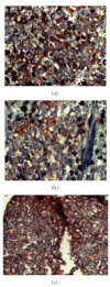
Primitive neuroectodermal tumor (PNET) of the chest wall or Askin's tumor is a rare neoplasm of chest wall. It most often affects children and adolescents and is a very rare tumor in adults. In this case report, we present an Askin's tumor occurred in a 73-year-old male. The patient was admitted with a history of 3-month lower back pain and cough. In computed tomography, there was a lesion with dimensions of 70 x 40 x 65 mm in the superior segment of the lower lobe of the left lung. Positron emission tomography/computed tomography with 18F-flourodeoxyglucose revealed a pleural-based tumor in the left lung with a maximum standardized uptake value of 4.36. No distant or lymph node metastases were present. The patient had gone through surgery, and wedge resection of the superior segment of left lobe and partial resection of the ipsilateral ribs were performed. Pathology report with immunocytochemistry was consistent with PNET and the patient received chemotherapy after that.
Figures


References
-
- Harimaya K, Oda Y, Matsuda S, Tanaka K, Chuman H, Iwamoto Y. Primitive neuroectodermal tumor and extraskeletal Ewing sarcoma arising primarily around the spinal column: report of four cases and a review of the literature. Spine. 2003;28(19):E408–E412. - PubMed
-
- Askin FB, Rosai J, Sibley RK, Dehner LP, McAlister WH. Malignant small cell tumor of the thoracopulmonary region in childhood. A distinctive clinicopathological entity of uncertain histogenesis. Cancer. 1979;43(6):2438–2451. - PubMed
-
- Cabezali R, Lozano R, Bustamante E, et al. Askin's tumor of the chest wall: a case report in an adult. Journal of Thoracic and Cardiovascular Surgery. 1994;107(3):960–962. - PubMed
-
- Burge HJ, Novotny DB, Schiebler ML, Delany DJ, McCartney WH. MRI of Askin's tumor. Case report at 1.5 T. Chest. 1990;97(5):1252–1254. - PubMed
-
- Ravaux S, Bousqoet JC, Vancina S. Askin's tumor in a 67-year-old man with cancer of the prostate. X-ray computed tomography aspects. Journal de Radiologie. 1990;71(3):233–236. - PubMed
Publication types
LinkOut - more resources
Full Text Sources

