. 2010 Mar;11(1):9-19.
doi: 10.1007/s10969-009-9075-x. Epub 2010 Jan 5.

Characterization of proteins with wide-angle X-ray solution scattering (WAXS)

Affiliations

Characterization of proteins with wide-angle X-ray solution scattering (WAXS)

Lee Makowski. J Struct Funct Genomics. 2010 Mar.

Abstract

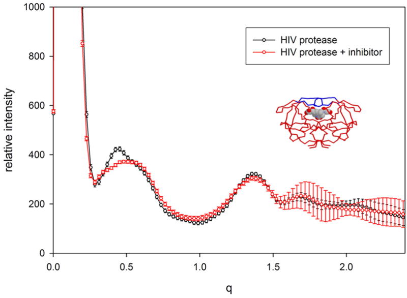
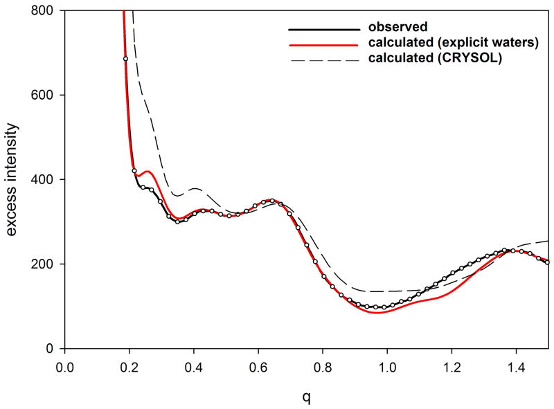
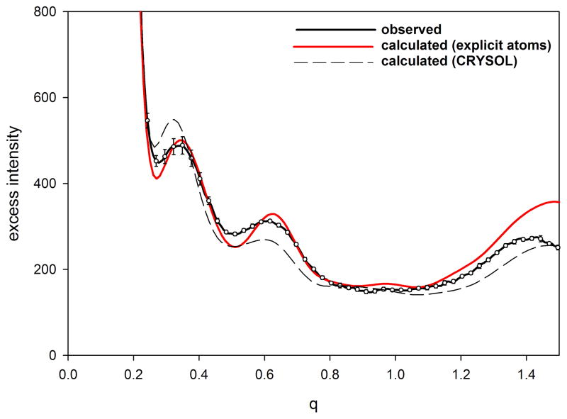
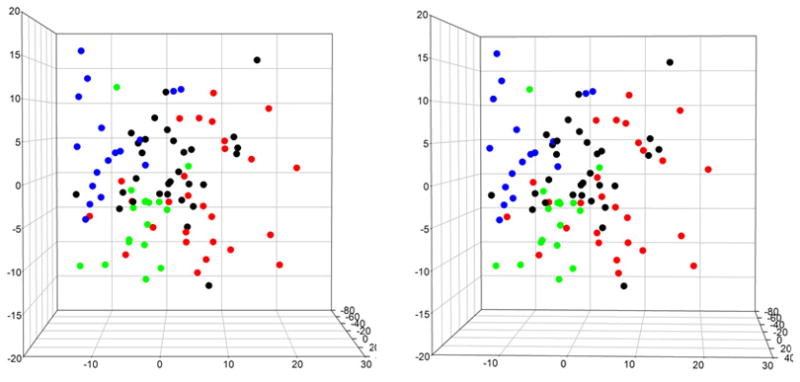
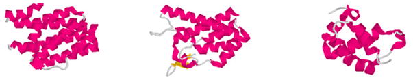
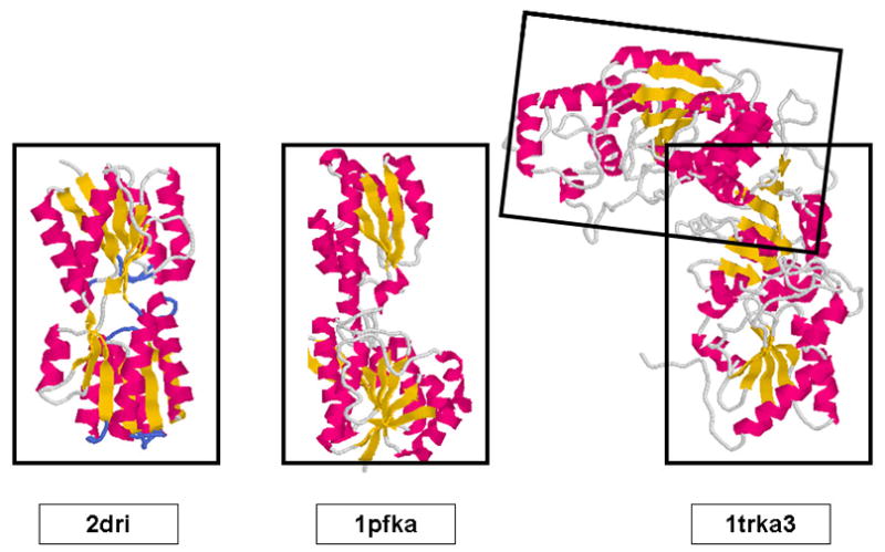
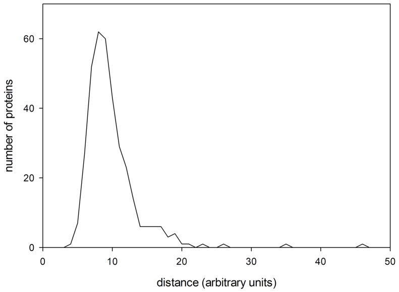
X-ray solution scattering in both the small-angle (SAXS) and wide-angle (WAXS) regimes is making an increasing impact on our understanding of biomolecular complexes. The accurate calculation of WAXS patterns from atomic coordinates has positioned the approach for rapid growth and integration with existing Structural Genomics efforts. WAXS data are sensitive to small structural changes in proteins; useful for calculation of the pair-distribution function at relatively high resolution; provides a means to characterize the breadth of the structural ensemble in solution; and can be used to identify proteins with similar folds. WAXS data are often used to test structural models, identify structural similarities and characterize structural changes. WAXS is highly complementary to crystallography and NMR. It holds great potential for the testing of structural models of proteins; identification of proteins that may exhibit novel folds; characterization of unfolded or natively disordered proteins; and detection of structural changes associated with protein function.


Figures

Figure 1
Figure 1
WAXS patterns from HIV protease (black) and HIV protease with the inhibitor MVT-101 bound (red). The differences between these two curves are statistically significant, being concentrated in the region q ~ 0.4 – 0.5, and reflecting the closing of the active site flaps in response to the binding of inhibitor. The inset is a rendering of the protease with the inhibitor represented as a space-filling model and the protein as a background tracing. In the apo form, the flaps (rendered blue in the inset) appear to exhibit substantial flexibility - movement that is required for entry of substrate (or inhibitor) into the active site. HIV protease was chemically synthesized and kindly provided by Vladimir Torbeev and Steve Kent (University of Chicago). Both samples had protein concentrations of 10 mg/ml. To enhance representation of the error bars, only 10% of the measured intensities are plotted.
Figure 2
Figure 2
Comparison of observed and calculated Iexcess from (a) equine myoglobin and (b) hen egg white lysozyme. Protein samples had a concentration of 20 mg/ml and a temperature of 20°C. Calculation was carried out with EXCESS using an explicit atomic representation of water (Park et al., 2009). Error bars represent the standard deviation of 10 exposures and cannot be seen in the data from myoglobin because they are smaller than the diameter of the circles representing the observations. To enhance representation of the error bars, only 10% of the measured intensities are plotted.
Figure 2
Figure 2
Comparison of observed and calculated Iexcess from (a) equine myoglobin and (b) hen egg white lysozyme. Protein samples had a concentration of 20 mg/ml and a temperature of 20°C. Calculation was carried out with EXCESS using an explicit atomic representation of water (Park et al., 2009). Error bars represent the standard deviation of 10 exposures and cannot be seen in the data from myoglobin because they are smaller than the diameter of the circles representing the observations. To enhance representation of the error bars, only 10% of the measured intensities are plotted.
Figure 3
Figure 3
Stereo pair showing the distribution of α (red), β (blue), α/β (green) and α+β (black) proteins in WAXS space, projected onto the coordinates corresponding to the 2nd, 3rd and 4th most significant eigenvectors.
Figure 4
Figure 4
Clustering in WAXS space. Renderings of 1hw1 (left) and two of its nearest neighbors in WAXS space. These three domains are members of the 498 distinct domains chosen to represent all of fold space (Hou et al., 2003). Nonetheless, their structures are clearly related. Other proteins exhibiting WAXS patterns that place them in the region of WAXS space can be shown to have closely related folds.
Figure 5
Figure 5
Comparison of the structure of 2dri with that of the A-chains of 1pfk and 1trk. Qualitatively, the structures of the domains of these three proteins appear to be very similar. 1trk consists of a pair of similar domains, each of which has similarities to 2dri.
Figure 6
Figure 6
Histogram of nearest neighbor distances between proteins representing different folds in the Pilot Database. On average, the nearest neighbors of proteins in this database are no more than 8–9 (arbitrary) units away. Proteins having the same folds are usually found closer to one another than that. Proteins more than 8–9 units from any protein of known structure may represent a novel fold.

References

    1. Baker D, Sali A. Protein structure prediction and structural genomics. Science. 2001;294:93–96. - PubMed
    1. Bardhan JP, Park S, Makowski L. SoftWAXS: A Computational Tool for Modeling Wide-X-ray Solution Scattering from Biomolecules. 2009. Submitted for publication. - PMC - PubMed
    1. Cammarata M, Levantino M, Schotte F, Anfinrud PA, Ewald F, Choi J, Cupane A, Wulff M, Ihee J. Tracking the structural dynamics of proteins in solution using time-resolved wide-angle X-ray scattering. Nature Methods. 2008;5:881–886. - PMC - PubMed
    1. Chacon P, Diaz JF, Moran F, Andreu JM. Reconstruction of protein form with x-ray solution scattering and a genetic algorithm. J Mol Biol. 2000;299:1289–1302. - PubMed
    1. Clarage JB, Romo T, Andrews BK, Pettitt BM, Phillips GN. A sampling problem in molecular dynamics simulations of macromolecules. Proc Natl Acad Sci. 1995;92:3288–3292. - PMC - PubMed

Publication types

LinkOut - more resources