

Tendon properties remain altered in a chronic rat rotator cuff model
- PMID: 20049569
- PMCID: PMC2865621
- DOI: 10.1007/s11999-009-1206-y
Tendon properties remain altered in a chronic rat rotator cuff model
Abstract
Background: Chronic rotator cuff tears are often associated with pain or poor function. In a rat with only a detached supraspinatus tendon, the tendon heals spontaneously which is inconsistent with how tears are believed to heal in humans.
Questions/purposes: We therefore asked whether a combined supraspinatus and infraspinatus detachment in the rat would fail to heal and result in a chronic injury in the supraspinatus tendon.
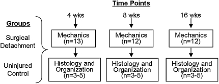
Methods: We acutely detached the supraspinatus and infraspinatus tendons in a rat model. At 4, 8, and 16 weeks post-detachment, biomechanical testing, collagen organization, and histological grading were evaluated for the detached supraspinatus and infraspinatus tendons and compared to controls.
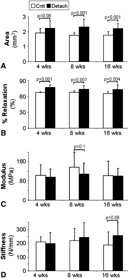
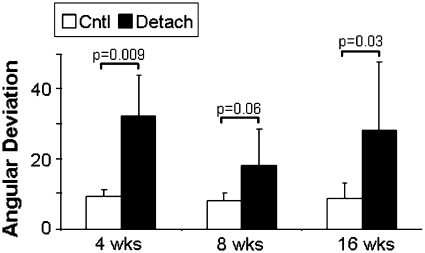
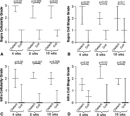
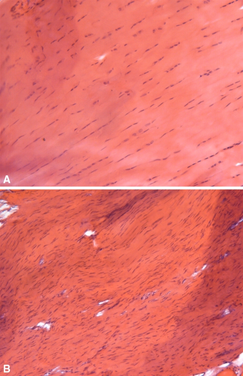
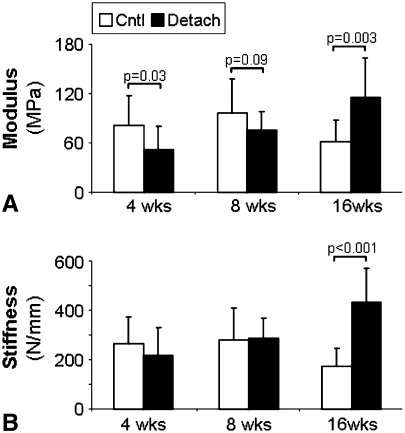
Results: In the detached supraspinatus tendon, area and percent relaxation were increased at all time points while the modulus and stiffness were similar to those of controls at 4 and 8 weeks. Collagen disorganization increased at late time points while cellularity increased and cells were more rounded in shape. In the detached infraspinatus tendon, area and percent relaxation were also increased at late time points. However, the modulus values initially decreased followed by an increase in both modulus and stiffness at 16 weeks compared to control. In the detached infraspinatus, we also observed a decrease in collagen organization at all time points and increased cellularity and a more rounded cell shape.
Conclusions: Due to the ongoing changes in mechanics, collagen organization and histology in the detached supraspinatus tendon compared to control animals at 16 weeks, this model may be useful for understanding the human chronic tendon tear.
Clinical relevance: This rat rotator cuff chronic model can be used to test hypotheses regarding injury and repair mechanisms that cannot be addressed in human patients or in cadaveric studies.
Figures
Similar articles
-
Rat rotator cuff tendon-to-bone healing properties are adversely affected by hypercholesterolemia.J Shoulder Elbow Surg. 2014 Jun;23(6):867-72. doi: 10.1016/j.jse.2013.08.018. Epub 2013 Dec 2. J Shoulder Elbow Surg. 2014. PMID: 24295837 Free PMC article.
-
Supraspinatus tendon organizational and mechanical properties in a chronic rotator cuff tear animal model.J Biomech. 2004 May;37(5):739-49. doi: 10.1016/j.jbiomech.2003.09.019. J Biomech. 2004. PMID: 15047003
-
After rotator cuff tears, the remaining (intact) tendons are mechanically altered.J Shoulder Elbow Surg. 2009 Jan-Feb;18(1):52-7. doi: 10.1016/j.jse.2008.07.003. J Shoulder Elbow Surg. 2009. PMID: 19095175 Free PMC article.
-
Histopathology of rotator cuff tears.Sports Med Arthrosc Rev. 2011 Sep;19(3):227-36. doi: 10.1097/JSA.0b013e318213bccb. Sports Med Arthrosc Rev. 2011. PMID: 21822106 Review.
-
Effect of anterior supraspinatus tendon partial-thickness tears on infraspinatus tendon strain through a range of joint rotation angles.J Shoulder Elbow Surg. 2010 Jun;19(4):617-23. doi: 10.1016/j.jse.2009.10.003. Epub 2010 Jan 15. J Shoulder Elbow Surg. 2010. PMID: 20080051 Free PMC article. Review.
Cited by
-
Effect of implantation site and injury condition on host response to human-derived fascia lata ECM in a rat model.J Orthop Res. 2012 Mar;30(3):461-7. doi: 10.1002/jor.21529. Epub 2011 Aug 19. J Orthop Res. 2012. PMID: 21858856 Free PMC article.
-
Comparison of rotator cuff muscle architecture between humans and other selected vertebrate species.J Exp Biol. 2014 Jan 15;217(Pt 2):261-73. doi: 10.1242/jeb.083923. Epub 2013 Sep 26. J Exp Biol. 2014. PMID: 24072803 Free PMC article.
-
Rat rotator cuff tendon-to-bone healing properties are adversely affected by hypercholesterolemia.J Shoulder Elbow Surg. 2014 Jun;23(6):867-72. doi: 10.1016/j.jse.2013.08.018. Epub 2013 Dec 2. J Shoulder Elbow Surg. 2014. PMID: 24295837 Free PMC article.
-
Scapular dyskinesis is detrimental to shoulder tendon properties and joint mechanics in a rat model.J Orthop Res. 2014 Nov;32(11):1436-43. doi: 10.1002/jor.22693. Epub 2014 Jul 28. J Orthop Res. 2014. PMID: 25070580 Free PMC article.
-
The rat as a novel model for chronic rotator cuff injuries.Sci Rep. 2024 Mar 4;14(1):5344. doi: 10.1038/s41598-024-55281-5. Sci Rep. 2024. PMID: 38438458 Free PMC article.
References
-
- Bartolozzi A, Andreychik D, Ahmad S. Determinants of outcome in the treatment of rotator cuff disease. Clin Orthop Relat Res. 1994:90–97. - PubMed
-
- Bjorkenheim JM, Paavolainen P, Ahovuo J, Slatis P. Surgical repair of the rotator cuff and surrounding tissues. Factors influencing the results. Clin Orthop Relat Res. 1988:148–153. - PubMed
Publication types
MeSH terms
Substances
Grants and funding
LinkOut - more resources
Full Text Sources
Other Literature Sources
Medical
