

Inflammation and EMT: an alliance towards organ fibrosis and cancer progression
- PMID: 20049734
- PMCID: PMC3378143
- DOI: 10.1002/emmm.200900043
Inflammation and EMT: an alliance towards organ fibrosis and cancer progression
Abstract
Recent advances in our understanding of the molecular pathways that govern the association of inflammation with organ fibrosis and cancer point to the epithelial to mesenchymal transition (EMT) as the common link in the progression of these devastating diseases. The EMT is a crucial process in the development of different tissues in the embryo and its reactivation in the adult may be regarded as a physiological attempt to control inflammatory responses and to 'heal' damaged tissue. However, in pathological contexts such as in tumours or during the development of organ fibrosis, this healing response adopts a sinister nature, steering these diseases towards metastasis and organ failure. Importantly, the chronic inflammatory microenvironment common to fibrotic and cancer cells emerges as a decisive factor in the induction of the pathological EMT.
Figures
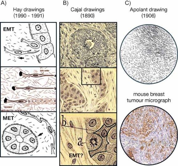
Betty Hay coined the term ‘epithelial to mesenchymal transformation’ (Hay, 1968) and described the transient nature of this process, as well as the reversion to the epithelial character (MET, Hay, 1991). Modified from Acloque et al (2008).
More than 100 years ago Santiago Ramón y Cajal drew and described the morphological appearance of breast carcinoma so accurately that we can find what we believe to be the first description of EMT (see text). Drawings are adapted from figs. 53 and 48 in Ramon y Cajal (1890). Note the morphology of the cell highlighted as ‘b’.
At the turn of the 20th century, Hugo Apolant also clearly represented the morphology of EMT-type mouse mammary tumours (Apolant, 1906). The micrograph shows the distribution of cytokeratin 8/18 in both the epithelial and spindle cell populations in a mouse mammary tumour diagnosed as EMT-type. This picture is courtesy of Dr P. Damonte and Dr R. Cardiff, Univ. California at Davis.
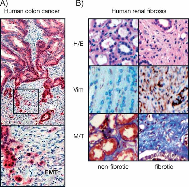
Immunohistochemical staining of a colorectal carcinoma for β-catenin in red and nuclear counterstaining in blue. The central area of the tumour exhibits polarized epithelial tumour cells lacking nuclear β-catenin, while cells at the invasive front undergo EMT and show nuclear β-catenin (Brabletz et al, 2001). Picture from Dr Thomas Brabletz, Univ. Freiburg (Germany).
Histological sections showing the halmarks of EMT in the medulla of fibrotic kidney from patients subjected to nephrectomy. Sections are stained with haematoxylin–eosin (H/E) to better appreciate cell morphology and the disappearance of the tubular structures in fibrosis, vimentin expression showing mesenchymal cells (brown, Vim) and fibrotic deposits revealed by the blue Masson–Trichome staining (M/T) (Boutet et al, 2006).
References
-
- Adorno M, Cordenonsi M, Montagner M, Dupont S, Wong C, Hann B, Solari A, Bobisse S, Rondina MB, Guzzardo V, et al. A mutnat-p53/Smad complex opposes p63 to empower TGFβ-induced metastasis. Cell. 2009;177:87–98. - PubMed
-
- Apolant H. Die epithelialen Geschwülste der Maus. Arb Königl Inst Exp Ther. 1906;1:7–62.
-
- Baan C, van Gelder T, Peeters A, Mol W, Niesters H, Weimar W, IJzermans J. Living kidney donors and hypoxia-inducible factor-1alpha. Transplantation. 2003;75:570–571. - PubMed
Publication types
MeSH terms
Substances
LinkOut - more resources
Full Text Sources
Other Literature Sources
