. 2009 Nov;1(8-9):381-91.
doi: 10.1002/emmm.200900045.

Molecular aging and rejuvenation of human muscle stem cells

Affiliations

Molecular aging and rejuvenation of human muscle stem cells

Morgan E Carlson et al. EMBO Mol Med. 2009 Nov.

Abstract

Very little remains known about the regulation of human organ stem cells (in general, and during the aging process), and most previous data were collected in short-lived rodents. We examined whether stem cell aging in rodents could be extrapolated to genetically and environmentally variable humans. Our findings establish key evolutionarily conserved mechanisms of human stem cell aging. We find that satellite cells are maintained in aged human skeletal muscle, but fail to activate in response to muscle attrition, due to diminished activation of Notch compounded by elevated transforming growth factor beta (TGF-beta)/phospho Smad3 (pSmad3). Furthermore, this work reveals that mitogen-activated protein kinase (MAPK)/phosphate extracellular signal-regulated kinase (pERK) signalling declines in human muscle with age, and is important for activating Notch in human muscle stem cells. This molecular understanding, combined with data that human satellite cells remain intrinsically young, introduced novel therapeutic targets. Indeed, activation of MAPK/Notch restored 'youthful' myogenic responses to satellite cells from 70-year-old humans, rendering them similar to cells from 20-year-old humans. These findings strongly suggest that aging of human muscle maintenance and repair can be reversed by 'youthful' calibration of specific molecular pathways.


Figures

Figure 1
Figure 1. Immobility-induced muscle atrophy causes an age-specific increase in degeneration and lack of myogenic recovery
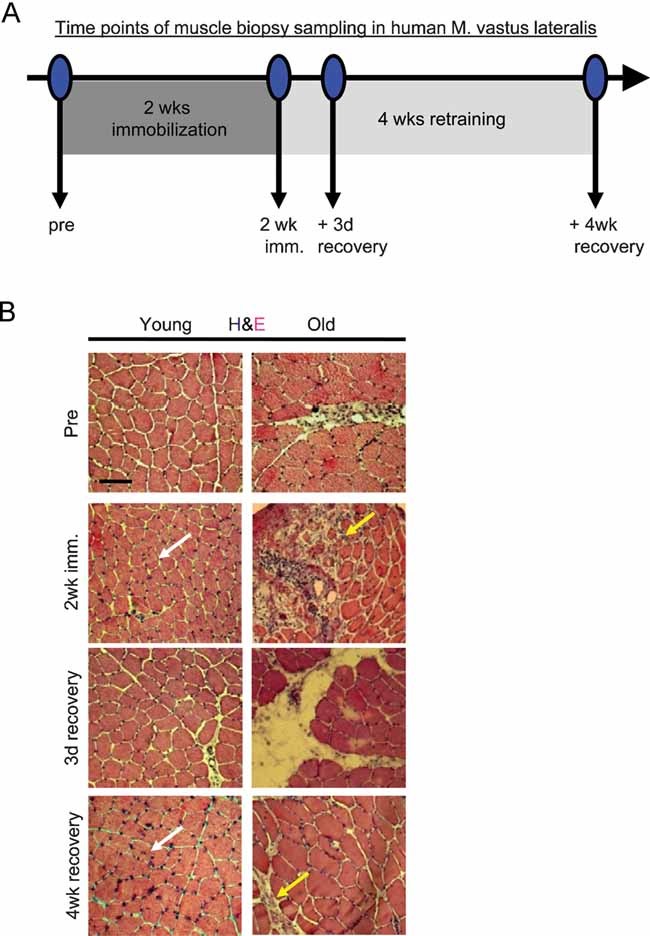
  1. Scheme of experimental setup, as described in text.

  2. Muscle histology from resting state (pre), immobility-atrophy (2 week imm.) and loading-recovery (3 day recovery, 4 week recovery) was analysed by haematoxylin and eosin (H & E) of 10-µm skeletal muscle cryosections. During immobilization phase of the study, areas of severe degeneration and scar tissue formation were evident in old muscles (yellow arrows) versus healthy maintenance of young immobilized muscles (white arrows). Scale bar = 100 µm. n = 10.

Figure 2
Figure 2. Pronounced lack of myogenic cell expansion is detected in old human muscle that undergoes exercise after immobility
A. Skeletal muscle cryosections were immunostained for Pax7, M-Cadherin and NCAM (green). Laminin immuno-detection is shown in red and Hoechst labels nuclei (blue). Sublaminar mono-nucleated cells expressing these markers were identified in association with both young and old myofibres. B. The total numbers of Pax7, NCAM and M-Cadherin+ satellite cell nuclei were quantified per 10 mm2 for both young and old muscles. *P ≤ 0.05 (old compared to young at ‘pre’ condition). C. Total number of Pax7+ nuclei were analysed at the pre and 2 week (resting and immobilization phase) and the 2 week + 3 day and 4 week time points (regeneration phase). *, P ≤ 0.05 (young compared to old at 2 week + 3 day). D, E.Western blotting for Pax7 was performed on whole muscle protein isolates (quantified in E), using actin as a loading control. *P ≤ 0.05, old compared with young. Data are means ± s.d. n = 10 for immunostained cryosections, n = 6 for Western blot analysis.
Figure 3
Figure 3. Notch activation and Delta upregulation is diminished in regenerating old human skeletal muscle
A. Cryosections were analysed by immunostaining for co-expression of nuclear Pax7+/active Notch in resident satellite cells. B. Delta (red) and dystrophin (green) immuno-detection is shown for 10 µm skeletal muscle cryosections. Hoechst labels nuclei (blue). C, D. Western blot of Notch and Delta levels on whole muscle protein isolates for 2 week + 3 day; quantified in D. *P ≤ 0.05, old compared with young for both Notch and Delta. E. Quantification of Notch/Pax7 double-positive myofibre-associated cells from cryosections. Data are means ± s.d., n = 10–15 for immunodetection of cryosections. n = 6 for Western blotting analysis. As compared to young tissue, in old muscle loaded after immobility, there is significant down-regulation of Delta, active Notch and decline in numbers of myofibre-associated myogenic cells that co-express Pax7 and active Notch.
Figure 4
Figure 4. Mechanisms of muscle stem cell aging are conserved between mice and humans, with respect to TGF-β signalling imbalance
Immunodetection of: A. TGF-β (green) and laminin (red), B. P-Smad3 (green) and laminin (red) is shown for 10 µm skeletal muscle cryosections. Hoechst labels nuclei (blue). C, D. Western blotting for TGF-β, P-Smad3, p15, p21, p16 and p27 from whole muscle protein lysates; quantified in D. Actin was used as loading control. *P ≤ 0.05, old compared with young. Significant age-specific elevation of TGF-β/pSmad and of CDK inhibitors, p15 and p21 was detected in old muscle as compared to young. E, F. Activated satellite cells were cultured for 24 h in OPTI-MEM containing age-matched human sera in the presence of 25 ng/ml recombinant TGF-β1. Myogenic responses were analysed and quantified F, based on the co-expression of desmin/BrdU. Data are means ± s.d., n = 10–15 for immunodetection of cryosections, n = 6 for Western blotting analysis, n = 6 for myogenic culture experiments.
Figure 5
Figure 5. MAPK signalling strength becomes diminished in old human muscle, and is causal for Notch activation and myogenic properties of human satellite cells
A, B. Western blot on whole muscle protein isolates was performed for P-Erk and Erk, quantified in B where *, P ≤ 0.05, old compared with young. Similar levels of Erk, but lower levels of pErk were detected in old muscle as compared to young. C, D. Western blot of Notch, Delta, P-Erk and Erk was performed (quantified in D), following 24 h exogenous addition of 10 ng/ml FGF or 10 µM MEK inhibitor (MEKi) to isolated human satellite cell cultures *, **P ≤ 0.05, +FGF compared to Control and +MEKi compared to Control. Experimental attenuation of MAPK decreases levels of Delta and active Notch, while induction of MAPK up-regulates Delta and active Notch. E, F. Activated satellite cell, cultured as in C, were analysed for myogenic responses and quantified F, based on the co-expression of desmin/BrdU. Data are means ± s.d., n = 6 for all panels indicated. Myogenic responses of both young and old human satellite cells were enhanced by forced activation of MAPK and were diminished by attenuation of MAPK.
Figure 6
Figure 6. Notch is a necessary and sufficient molecular determinant of human myogenic responses in vitro, which rescues productive regeneration in the presence of aged sera and attenuates expression levels of p15 and p21 in human satellite cell cultures
A, B. Young and old satellite cells were isolated and cultured in the presence of young or old sera, with or without experimentally induced Notch activation (immobilized Delta ligand for old cells) or Notch inhibition (treatment with GSI for young cells). After seven days of culture, cells were fixed and analysed for myogenic responses (generation of desmin/BrdU co-stained cells). Quantification is shown in B. *P ≤ 0.05 (Y + Y sera compared to O + O sera and Y + Y sera/GSI, O + O sera/Delta compared to O + O sera). n = 6. Representative immunostaining is depicted in A; desmin (green) BrdU (red), Hoechst (blue) labels nuclei. Activation of Notch is required for myogenic properties of young satellite cells and rescues myogenic potential of old human satellite cells even in the presence of aged human sera. Young satellite cells did not have significantly higher myogenic potential when Notch was experimentally activated, as compared to control young cells, suggesting that young myogenic responses are at the optimal high (not shown). C, D. Treated cells were analysed for the expression of p15 and p21 by Western blot; quantified in D. Actin serves as loading control. *P ≤ 0.05, Y + GSI compared to Y + Y and O + Delta compared to O + O. Data are means ± s.d., n = 6 for immunodetection assays and Western blot analyses. Forced activation of Notch reduces levels of p15 and p21, while experimental attenuation of Notch activity increases the levels of these CDK inhibitors in satellite cells.

References

    1. Bergstrom J, Alvestrand A, Furst P, Hultman E, Sahlin K, Vinnars E, Widstrom A. Influence of severe potassium depletion and subsequent repletion with potassium on muscle electrolytes, metabolites and amino acids in man. Clin Sci Mol Med Suppl. 1976;51:589–599. - PubMed
    1. Brack AS, Conboy IM, Conboy MJ, Shen J, Rando TA. A temporal switch from notch to Wnt signaling in muscle stem cells is necessary for normal adult myogenesis. Cell Stem Cell. 2008;2:50–59. - PubMed
    1. Brooke MH, Kaiser KK. Muscle fiber types: how many and what kind? Arch Neurol. 1970;23:369–379. - PubMed
    1. Brown M, Hasser EM. Differential effects of reduced muscle use (hindlimb unweighting) on skeletal muscle with aging. Aging (Milano) 1996;8:99–105. - PubMed
    1. Carlson ME, Conboy IM. Loss of stem cell regenerative capacity within aged niches. Aging Cell. 2007;6:371–382. - PMC - PubMed

Publication types

MeSH terms

Substances