

Review
doi: 10.1002/wnan.40.
Top-down particle fabrication: control of size and shape for diagnostic imaging and drug delivery
Affiliations
- PMID: 20049805
- PMCID: PMC2804992
- DOI: 10.1002/wnan.40
Item in Clipboard
Review
Top-down particle fabrication: control of size and shape for diagnostic imaging and drug delivery
Wiley Interdiscip Rev Nanomed Nanobiotechnol.
2009 Jul-Aug.
Abstract
This review discusses rational design of particles for use as therapeutic vectors and diagnostic imaging agent carriers. The emerging importance of both particle size and shape is considered, and the adaptation and modification of soft lithography methods to produce nanoparticles are highlighted. To this end, studies utilizing particles made via a process called Particle Replication In Non-wetting Templates are discussed. In addition, insights gained into therapeutic cargo and imaging agent delivery from related types of polymer-based carriers are considered.
(c) 2009 John Wiley & Sons, Inc.
Figures
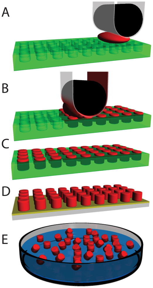
Schematic representation of the PRINT process. a.) Empty mold (green), high surface energy polymer sheet (clear), roller (black) is brought into contact with the particle precursor solution (red) and the mold; b.) Roller evenly distributes particle precursor solution into the cavities of mold. Excess particle precursor solution is wicked away by the high surface energy polymer sheet; c.) Particles are cured in the mold; d.) Particles are removed from the mold; e.) Particles are collected or harvested using a number of different film based techniques and ultimately are dispersed in solution.
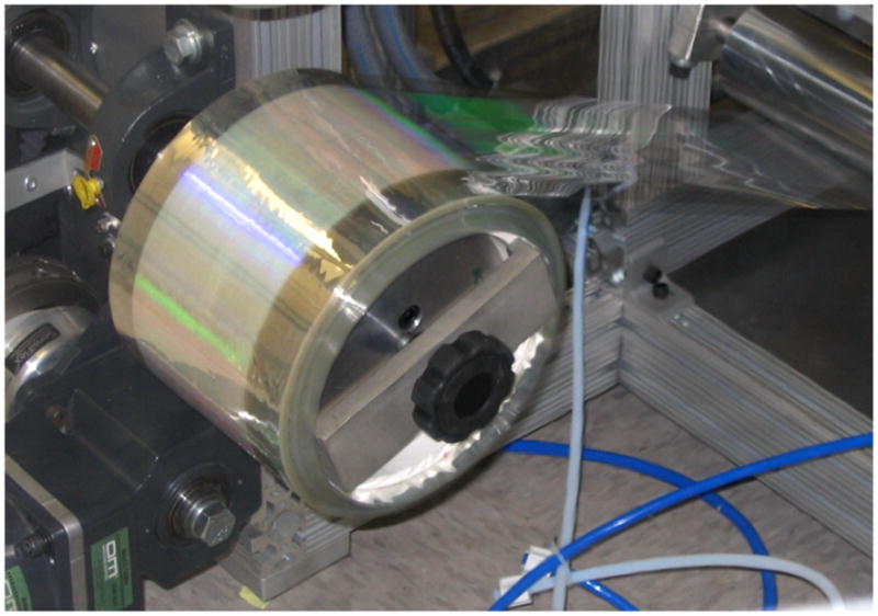
Continuous thin mold manufacturing for the PRINT process. Patterned surface can be seen in green. Reprinted with permission from Liquidia Technologies.[71]
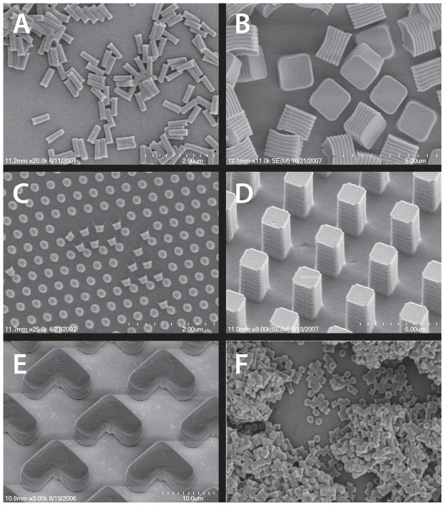
SEM images of particles of various sizes, shapes, and compositions prepared via the PRINT process: (A) Hydrogel rods containing antisense oligonucleotide; (B) crosslinked degradable matrix cubes containing Doxorubicin HCl; (C) Abraxane™ harvested onto medical adhesive; (D) Insulin particles harvested onto a medical adhesive; (E) Hydrogel “boomerangs” containing 15 wt% iron oxide; (F) Hydrogel cylinders containing 10 wt% Omniscan™. Reprinted with permission from references (B) [47];(C) [48]; (D) [48]; (E) [46].
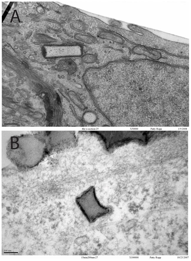
TEM showing HeLa cell internalization of 150 nm × 450 nm (top) or 200 nm × 200 nm (bottom) cylindrical particles fabricated via the PRINT process. Reprinted with permission from [44].
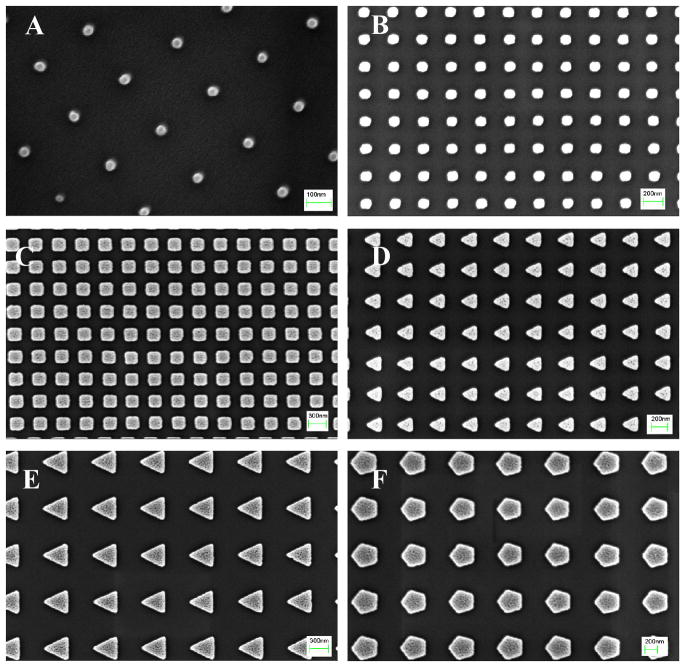
SEM images of S-FIL imprinted (100% w/v, MW 3400) PEGDA nanoparticles: (A) 50 nm squares (scale bar = 100 nm), (B) 100 nm squares (scale bar = 200 nm), (C) 200 nm squares (scale bar = 300 nm), (D) 200 nm triangles (scale bar = 200 nm), (E) 400 nm triangles (scale bar = 300 nm), and (F) 400 nm pentagonal particles (scale bar = 200 nm). Reprinted with permission from [68]
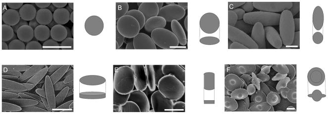
a) Scanning electron micrographs and 3D illustrations of PS particles created for phagocytosis experiments. (A) Spheres. (B) Oblate ellipsoids (13%). (C) Prolate ellipsoids (7%). (D) Elliptical disks (9%). (E) Rectangular disks (5%). (F) UFOs (12%). Particles are monodispersed with average standard deviations of measured dimensions for each shape listed in parentheses. A portion of this variation is due to 2–5%standard deviation in the diameter of spheres used as starting materials. (Scale bars: 5 m.) Reprinted with permission from [62]
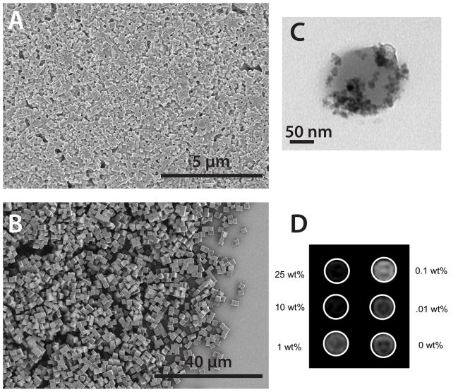
Magneto-polymer composite polymer particles of well defined size and shape prepared via the PRINT process: (A) SEM image of 200 nm × 200 nm cylinders, (B) SEM image of 2×2×2 micron cubes (C) TEM of biocompatible 200 × 200 nm particle containing 15 wt% PEG-silane coated iron oxide nanocrystals, and (D) T2 phantom study of iron oxide containing particles in agarose gel. Equimolar concentrations of PRINT particles with increasing iron oxide loading.
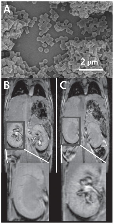
T1 weighted MR images a bolus injection of 200 × 200 nm cylindrical particles (a) just before (b) and 60 minutes post (c) injection.
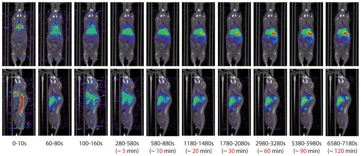
MicroPET imaging with 64Cu-DOTA PRINT particles. Time resolved PET images consisting of a two hour dynamic scan. The PET/CT images are overlayed. Mouse was injected with 136.2 μCi of 64Cu-labeled DOTA-nanoparticle. Both the coronal view (top), and sagittal view (bottom) are presented. Reprinted with permission from [45]
Similar articles
-
Engineering DNA scaffolds for delivery of anticancer therapeutics.Biomater Sci. 2015 Jul;3(7):1018-24. doi: 10.1039/c4bm00459k. Epub 2015 Feb 25. Biomater Sci. 2015. PMID: 26221936
-
Advantages of nanotechnology-based Chinese herb drugs on biological activities.Curr Drug Metab. 2009 Oct;10(8):905-13. doi: 10.2174/138920009790274603. Curr Drug Metab. 2009. PMID: 20214585 Review.
-
Nanoparticles and their applications in cell and molecular biology.Integr Biol (Camb). 2014 Jan;6(1):9-26. doi: 10.1039/c3ib40165k. Integr Biol (Camb). 2014. PMID: 24104563 Free PMC article. Review.
-
Particle shape effects in vitro and in vivo.Front Biosci (Schol Ed). 2012 Jun 1;4(4):1344-53. doi: 10.2741/s336. Front Biosci (Schol Ed). 2012. PMID: 22652876 Review.
-
Mesoporous silica nanoparticles improve magnetic labeling efficiency in human stem cells.Small. 2008 May;4(5):619-26. doi: 10.1002/smll.200700493. Small. 2008. PMID: 18491363
Cited by
-
Chemistry with spatial control using particles and streams().RSC Adv. 2012 Oct 28;2(26):9707-9726. doi: 10.1039/C2RA20337E. RSC Adv. 2012. PMID: 23145348 Free PMC article.
-
Analysis of the murine immune response to pulmonary delivery of precisely fabricated nano- and microscale particles.PLoS One. 2013 Apr 12;8(4):e62115. doi: 10.1371/journal.pone.0062115. Print 2013. PLoS One. 2013. PMID: 23593509 Free PMC article.
-
Shaping cancer nanomedicine: the effect of particle shape on the in vivo journey of nanoparticles.Nanomedicine (Lond). 2014 Jan;9(1):121-34. doi: 10.2217/nnm.13.191. Nanomedicine (Lond). 2014. PMID: 24354814 Free PMC article. Review.
-
Designer nanoparticles: incorporating size, shape and triggered release into nanoscale drug carriers.Expert Opin Drug Deliv. 2010 Apr;7(4):479-95. doi: 10.1517/17425240903579971. Expert Opin Drug Deliv. 2010. PMID: 20331355 Free PMC article. Review.
-
Effect of aspect ratio and deformability on nanoparticle extravasation through nanopores.Langmuir. 2012 Jun 12;28(23):8773-81. doi: 10.1021/la301279v. Epub 2012 May 29. Langmuir. 2012. PMID: 22612428 Free PMC article.
References
-
- Wong SY, Pelet JM, Putnam D. Polymer Systems for Gene Delivery - Past, Present, and Future. Prog Polym Sci. 2007;32:799–837.
-
- Zamboni WC. Concept and Clinical Evaluation of Carrier-Mediated Anticancer Agents. The Oncologist. 2008;13:248–260. - PubMed
-
- Nie S, Xing Y, Kim GJ, Simons JW. Nanotechnology: Applications in Cancer. Annu Rev Biomed Eng. 2007;9:257–288. - PubMed
-
- Cuchelkar V, Kopeček J. Polymer-Drug Conjugates. In: Uchegbu IF, Schaetzlain AG, editors. Polymers in Drug Delivery. Boca Raton, FL: CRC Press LLC; 2006. pp. 155–182.
-
- Duncan R. The Dawning Era of Polymer Therapeutics. Nature Reviews Drug Discovery. 2003;2:347–360. - PubMed
Publication types
MeSH terms
Substances
Grants and funding
LinkOut - more resources
Full Text Sources
Other Literature Sources
Medical
