

Biopersistence and potential adverse health impacts of fibrous nanomaterials: what have we learned from asbestos?
- PMID: 20049814
- PMCID: PMC2864601
- DOI: 10.1002/wnan.41
Biopersistence and potential adverse health impacts of fibrous nanomaterials: what have we learned from asbestos?
Abstract
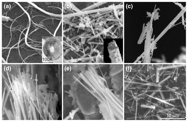
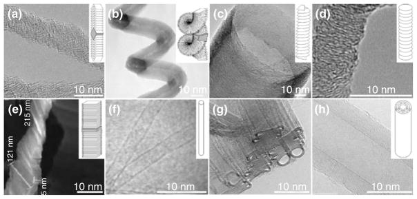
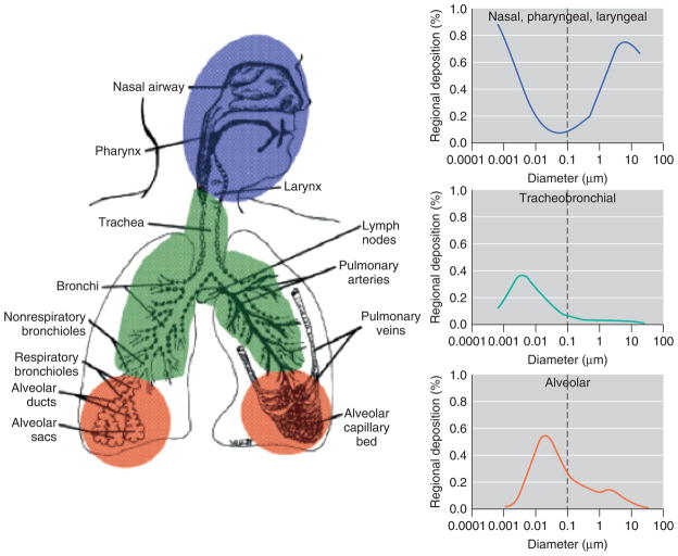
Human diseases associated with exposure to asbestos fibers include pleural fibrosis and plaques, pulmonary fibrosis (asbestosis), lung cancer, and diffuse malignant mesothelioma. The critical determinants of fiber bioactivity and toxicity include not only fiber dimensions, but also shape, surface reactivity, crystallinity, chemical composition, and presence of transition metals. Depending on their size and dimensions, inhaled fibers can penetrate the respiratory tract to the distal airways and into the alveolar spaces. Fibers can be cleared by several mechanisms, including the mucociliary escalator, engulfment, and removal by macrophages, or through splitting and chemical modification. Biopersistence of long asbestos fibers can lead to inflammation, granuloma formation, fibrosis, and cancer. Exposure to synthetic carbon nanomaterials, including carbon nanofibers and carbon nanotubes (CNTs), is considered a potential health hazard because of their physical similarities with asbestos fibers. Respiratory exposure to CNTs can produce an inflammatory response, diffuse interstitial fibrosis, and formation of fibrotic granulomas similar to that observed in asbestos-exposed animals and humans. Given the known cytotoxic and carcinogenic properties of asbestos fibers, toxicity of fibrous nanomaterials is a topic of intense study. The mechanisms of nanomaterial toxicity remain to be fully elucidated, but recent evidence suggests points of similarity with asbestos fibers, including a role for generation of reactive oxygen species, oxidative stress, and genotoxicity. Considering the rapid increase in production and use of fibrous nanomaterials, it is imperative to gain a thorough understanding of their biologic activity to avoid the human health catastrophe that has resulted from widespread use of asbestos fibers.
Copyright (c) 2009 John Wiley & Sons, Inc.
Figures
Similar articles
-
The asbestos-carbon nanotube analogy: An update.Toxicol Appl Pharmacol. 2018 Dec 15;361:68-80. doi: 10.1016/j.taap.2018.06.027. Epub 2018 Jun 28. Toxicol Appl Pharmacol. 2018. PMID: 29960000 Free PMC article. Review.
-
Pulmonary toxicity of carbon nanotubes and asbestos - similarities and differences.Adv Drug Deliv Rev. 2013 Dec;65(15):2078-86. doi: 10.1016/j.addr.2013.07.014. Epub 2013 Jul 27. Adv Drug Deliv Rev. 2013. PMID: 23899865 Review.
-
Toxicokinetics and effects of fibrous and nonfibrous particles.Inhal Toxicol. 2002 Jan;14(1):29-56. doi: 10.1080/089583701753338622. Inhal Toxicol. 2002. PMID: 12122559 Review.
-
A 3-dimensional in vitro model of epithelioid granulomas induced by high aspect ratio nanomaterials.Part Fibre Toxicol. 2011 May 18;8:17. doi: 10.1186/1743-8977-8-17. Part Fibre Toxicol. 2011. PMID: 21592387 Free PMC article.
-
Comparison of Calidria chrysotile asbestos to pure tremolite: final results of the inhalation biopersistence and histopathology examination following short-term exposure.Inhal Toxicol. 2005 Aug;17(9):427-49. doi: 10.1080/08958370591002012. Inhal Toxicol. 2005. PMID: 16020039
Cited by
-
Synchrotron soft X-ray imaging and fluorescence microscopy reveal novel features of asbestos body morphology and composition in human lung tissues.Part Fibre Toxicol. 2011 Feb 7;8(1):7. doi: 10.1186/1743-8977-8-7. Part Fibre Toxicol. 2011. PMID: 21299853 Free PMC article.
-
Attenuation of oxidative stress and chromosomal aberrations in cultured macrophages and pulmonary cells following self-sustained high temperature synthesis of asbestos.Sci Rep. 2020 May 22;10(1):8581. doi: 10.1038/s41598-020-65620-x. Sci Rep. 2020. PMID: 32444646 Free PMC article.
-
Lung macrophages "digest" carbon nanotubes using a superoxide/peroxynitrite oxidative pathway.ACS Nano. 2014 Jun 24;8(6):5610-21. doi: 10.1021/nn406484b. Epub 2014 Jun 4. ACS Nano. 2014. PMID: 24871084 Free PMC article.
-
Carbon Nanotube Emissions from Arc Discharge Production: Classification of Particle Types with Electron Microscopy and Comparison with Direct Reading Techniques.Ann Occup Hyg. 2016 May;60(4):493-512. doi: 10.1093/annhyg/mev094. Epub 2016 Jan 8. Ann Occup Hyg. 2016. PMID: 26748380 Free PMC article.
-
Animal models of fibrotic lung disease.Am J Respir Cell Mol Biol. 2013 Aug;49(2):167-79. doi: 10.1165/rcmb.2013-0094TR. Am J Respir Cell Mol Biol. 2013. PMID: 23526222 Free PMC article. Review.
References
-
- Kroto H, Heath J, O’Brien S, Curl R, Smalley R. C60:Buckminsterfullerene. Nature. 1985;318(14):162–163.
-
- Lam CW, James JT, McCluskey R, Arepalli S, Hunter RL. A review of carbon nanotube toxicity and assessment of potential occupational and environmental health risks. Crit Rev Toxicol. 2006;36(3):189–217. - PubMed
-
- Murr L, Soto K. TEM comparison of chrysotile (asbestos) nanotubes and carbon nanotubes. J Mater Sci. 2004;39:4941–4947.
-
- Stern S, Mcneil S. Nanotechnology safety concerns revisited. Toxicol Sci. 2008;101(1):4–21. - PubMed
Publication types
MeSH terms
Substances
Grants and funding
LinkOut - more resources
Full Text Sources
