

T-bet, a Th1 transcription factor regulates the expression of Tim-3
- PMID: 20049876
- PMCID: PMC2837127
- DOI: 10.1002/eji.200939842
T-bet, a Th1 transcription factor regulates the expression of Tim-3
Abstract
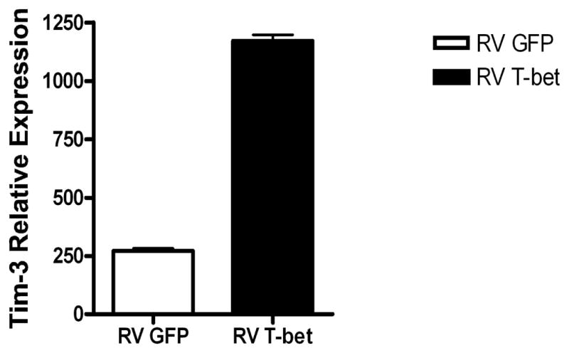
T-cell immunoglobulin, mucin domain-3 (Tim-3) is a membrane protein expressed at late stages of IFN-gamma secreting CD4(+) Th1 cell differentiation and constitutively on DC. Ligation of Tim-3 on Th1 cells terminates Th1 immune responses. In addition, Tim-3 plays a role in tolerance induction, although the mechanism by which this is accomplished has yet to be elucidated. While it is clear that Tim-3 plays an important role in the immune system, little is known regarding the molecular pathways that regulate Tim-3 expression. In the current study, we examine the role of Th1-associated transcription factors in regulating Tim-3 expression. Our experiments reveal that Tim-3 expression is regulated by the Th1-specific transcription factor T-bet. This introduces a novel paradigm into the generation of a Th1 response, whereby a transcription factor responsible for effector Th1 cell differentiation also increases the expression of a specific counter-regulatory molecule to ensure appropriate termination of pro-inflammatory Th1 immune responses.
Conflict of interest statement
VKK works with Telos Inc. which has funded part of this work. CAS and VKK hold a patent related to Tim-3 and Tim-3L. LHG has equity in and is on the corporate board of Bristol-Myers Squibb Company and is a paid consultant for HealthCare Ventures LLC Scientific Advisory Board and Mannkind Corporation. She has equity in MannKind Corporation and has filed patents that have been licensed by them.
Figures


References
-
- Monney L, Sabatos CA, Gaglia JL, Ryu A, Waldner H, Chernova T, Manning S, Greenfield EA, Coyle AJ, Sobel RA, Freeman GJ, Kuchroo VK. Th1-specific cell surface protein Tim-3 regulates macrophage activation and severity of an autoimmune disease. Nature. 2002;415:536–541. - PubMed
-
- Zhu C, Anderson AC, Schubart A, Xiong H, Imitola J, Khoury SJ, Zheng XX, Strom TB, Kuchroo VK. The Tim-3 ligand galectin-9 negatively regulates T helper type 1 immunity. Nat Immunol. 2005;6:1245–1252. - PubMed
-
- Sabatos CA, Chakravarti S, Cha E, Schubart A, Sanchez-Fueyo A, Zheng XX, Coyle AJ, Strom TB, Freeman GJ, Kuchroo VK. Interaction of Tim-3 and Tim-3 ligand regulates T helper type 1 responses and induction of peripheral tolerance. Nat Immunol. 2003;4:1102–1110. - PubMed
-
- Sanchez-Fueyo A, Tian J, Picarella D, Domenig C, Zheng XX, Sabatos CA, Manlongat N, Bender O, Kamradt T, Kuchroo VK, Gutierrez-Ramos JC, Coyle AJ, Strom TB. TIM-3 inhibits T helper type 1-mediated auto- and alloimmune responses and promotes immunological tolerance. Nat Immunol. 2003;4:1093–1101. - PubMed
-
- Anderson AC, Anderson DE, Bregoli L, Hastings WD, Kassam N, Lei C, Chandwaskar R, Karman J, Su EW, Hirashima M, Bruce JN, Kane LP, Kuchroo VK, Hafler DA. Promotion of tissue inflammation by the immune receptor Tim-3 expressed on innate immune cells. Science. 2007;318:1141–1143. - PubMed
Publication types
MeSH terms
Substances
Grants and funding
- R01 AI139671/AI/NIAID NIH HHS/United States
- NS38037/NS/NINDS NIH HHS/United States
- R01 CA112663/CA/NCI NIH HHS/United States
- K01 NS054096/NS/NINDS NIH HHS/United States
- NS30843/NS/NINDS NIH HHS/United States
- R37 NS030843/NS/NINDS NIH HHS/United States
- R01 NS035685/NS/NINDS NIH HHS/United States
- GM20927/GM/NIGMS NIH HHS/United States
- CA112663/CA/NCI NIH HHS/United States
- R29 NS030843/NS/NINDS NIH HHS/United States
- AI139671/AI/NIAID NIH HHS/United States
- R01 NS030843/NS/NINDS NIH HHS/United States
- F31 GM020927/GM/NIGMS NIH HHS/United States
- P01 NS038037/NS/NINDS NIH HHS/United States
- NS35685/NS/NINDS NIH HHS/United States
LinkOut - more resources
Full Text Sources
Other Literature Sources
Molecular Biology Databases
Research Materials
