. 2009 Dec;158(8):1874-83.
doi: 10.1111/j.1476-5381.2009.00475.x.

Multiple muscarinic pathways mediate the suppression of voltage-gated Ca2+ channels in mouse intestinal smooth muscle cells

Affiliations

Multiple muscarinic pathways mediate the suppression of voltage-gated Ca2+ channels in mouse intestinal smooth muscle cells

Yasuyuki Tanahashi et al. Br J Pharmacol. 2009 Dec.

Abstract

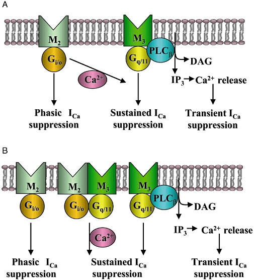
Background and purpose: Stimulation of muscarinic receptors in intestinal smooth muscle cells results in suppression of voltage-gated Ca2+ channel currents (I(Ca)). However, little is known about which receptor subtype(s) mediate this effect.

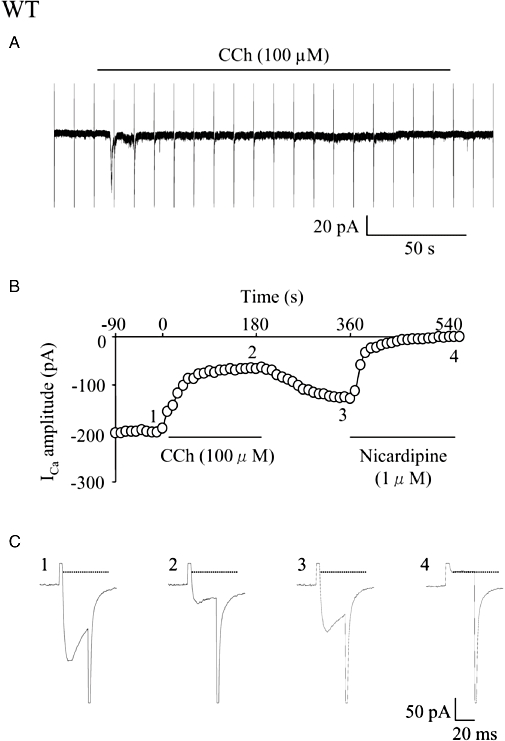
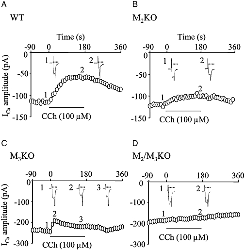
Experimental approach: The effect of carbachol on I(Ca) was studied in single intestinal myocytes from M2 or M3 muscarinic receptor knockout (KO) and wild-type (WT) mice.

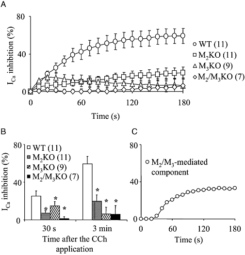
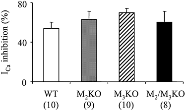
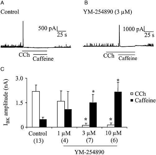
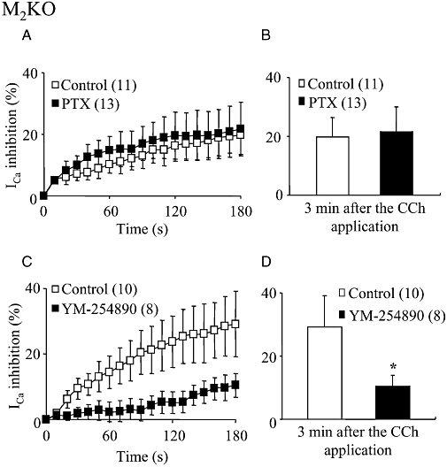
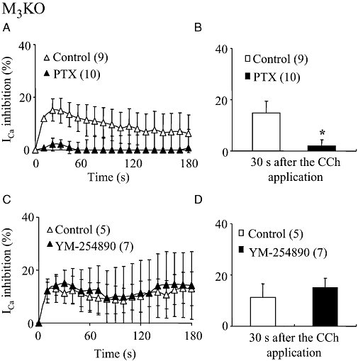
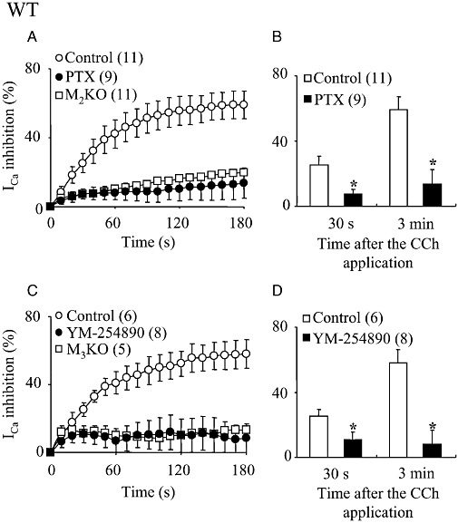
Key results: In M2KO cells, carbachol (100 microM) induced a sustained I(Ca) suppression as seen in WT cells. However, this suppression was significantly smaller than that seen in WT cells. Carbachol also suppressed I(Ca) in M3KO cells, but with a phasic time course. In M2/M3-double KO cells, carbachol had no effect on I(Ca). The extent of the suppression in WT cells was greater than the sum of the I(Ca) suppressions in M2KO and M3KO cells, indicating that it is not a simple mixture of M2 and M3 receptor responses. The G(i/o) inhibitor, Pertussis toxin, abolished the I(Ca) suppression in M3KO cells, but not in M2KO cells. In contrast, the G(q/11) inhibitor YM-254890 strongly inhibited only the I(Ca) suppression in M2KO cells. Suppression of I(Ca) in WT cells was markedly reduced by either Pertussis toxin or YM-254890.

Conclusion and implications: In intestinal myocytes, M2 receptors mediate a phasic I(Ca) suppression via G(i/o) proteins, while M3 receptors mediate a sustained I(Ca) suppression via G(q/11) proteins. In addition, another pathway that requires both M2/G(i/o) and M3/G(q/11) systems may be operative in inducing a sustained I(Ca) suppression.


Figures

Figure 3
Figure 3
Comparison of the inhibitory effect of carbachol (CCh) on the voltage-gated Ca2+ channel currents (ICa) in cells from wild-type (WT) and muscarinic receptor knockout (KO) mice. (A) Time courses of the change in the mean percentage suppression of ICa[the beginning of CCh (100 µM) application was taken as zero]. Each point indicates mean ± SEM. (B) The mean percentage suppression of ICa measured 30 s (four left-hand columns) and 3 min (four right-hand columns) after CCh application. Each column shows the mean with one SEM. In (A and B), the numbers of cells used are presented in parentheses. Asterisks in (B) represent statistically significant differences from the corresponding value in WT cells (P < 0.05). (C) An assumed sustained component of ICa suppression mediated by M2/M3 pathway. Each point was estimated by subtracting the average percentage suppression curve of ICa in both M2KO and M3KO cells from that in WT cells shown in (A).
Figure 1
Figure 1
Effects of carbachol (CCh) on the holding current and the voltage-gated Ca2+ channel currents (ICa) in a single longitudinal smooth muscle cell isolated from the small intestine of a wild-type (WT) mouse. ICa was elicited by a 30 ms step pulse to 0 mV from the holding potential of −60 mV at a frequency of 0.1 Hz. CCh (100 µM) was applied extracellularly during application of the depolarizing pulses. (A) CCh induced a cationic inward current (ICCh) consisting of an initial transient component followed by a sustained component. Every sharp current deflection shows overlapped ICa and the capacitive current. (B) Time course of the change in ICa amplitude (○) in the cell plotted against time (the beginning of CCh application was taken as zero). Points 1–4 in the graph correspond to actual ICa records (1–4) in (C). The amplitude of ICa was measured as the difference from the current level (interrupted line) obtained by the depolarizing pulse in the presence of nicardipine (1 µM). In the following figures, measurements and plots of ICa amplitude were made in the same way.
Figure 2
Figure 2
Effects of carbachol (CCh) on the voltage-gated Ca2+ channel currents (ICa) in cells isolated from muscarinic receptor knockout (KO) mice. (A–D) Time courses of the change in ICa amplitude in cells from wild-type (WT) (A), M2KO (B), M3KO (C) and M2/M3-double KO mice (D). Points 1–3 in each graph correspond to actual ICa records in insets 1–3.
Figure 4
Figure 4
Effect of guanosine 5′-(γ-thio) triphosphate tetralithium salt (GTP-γ-S) on the voltage-gated Ca2+ channel currents (ICa) in cells from wild-type (WT) and muscarinic receptor knockout (KO) mice. GTP-γ-S (200 µM) was applied intracellularly via patch pipettes. The mean percentage suppression of ICa measured 10 min after the establishment of whole-cell clamp configuration are shown. Each column represents the mean with one SEM. The numbers of cells used are shown in parentheses.
Figure 5
Figure 5
Effect of YM-254890 on BK current (IBK) in cells isolated from M2KO mice. CCh (100 µM)- or caffeine (10 mM)-induced Ca2+ release events were detected as IBK at the holding potential of 0 mV in M2KO cells. Cells were dialysed with a K+-rich pipette solution containing 0.3% DMSO (control) or YM-254890 (1–10 µM). (A and B) Typical current traces from a control cell (A) and YM-254890 (3 µM)-dialysed cell (B). (C) Summary of CCh- and caffeine- induced IBK in the presence or absence of YM-254890. The individual columns indicate the mean with one SEM. The numbers of cells used are shown in parentheses. Asterisks represent significant differences from the corresponding control values (P < 0.05). CCh, carbachol; DMSO, dimethyl sulphoxide; KO, knockout.
Figure 6
Figure 6
Effects of Pertussis toxin (PTX) and YM-254890 on the CCh-induced sustained ICa suppression in M2KO cells. (A) Time courses of the change in the mean percentage suppressions of ICa mediated by CCh (100 µM) in PTX-untreated (control) and -treated cells (the beginning of CCh application was taken as zero). (C) Time courses of the change in the mean percentage suppressions of ICa in 0.3% DMSO-dialysed (control) and YM-254890 (3 µM)-dialysed cells. Each point indicates mean ± SEM. The numbers of used cells are shown in parentheses. (B and D) Summary of the mean percentage suppression of ICa measured 3 min after the CCh application in control, PTX-treated (B) or YM-254890-dialysed cells (D). Each column shows the mean with one SEM. The asterisk in (D) represents a significant difference from the corresponding control value (P < 0.05). PTX had no effect on the sustained ICa suppression, but YM-254890 markedly diminished it. CCh, carbachol; DMSO, dimethyl sulphoxide; ICa, voltage-gated Ca2+ channel currents; KO, knockout.
Figure 7
Figure 7
Effects of PTX and YM-254890 on the CCh-induced phasic ICa suppression in M3KO cells. (A and C) Time courses of the change in the mean percentage suppression of ICa induced by CCh (100 µM) in PTX-treated (A) and YM-254890 (3 µM)-dialysed (C) M3KO cells plotted in the same way as in Figure 6. (B and D) Summary of the mean percentage suppression of ICa measured 30 s after CCh application in PTX-treated (B) and YM-254890-dialysed (D) cells. Asterisk in (B) represents significant difference from the corresponding control value (P < 0.05). PTX abolished the phasic ICa suppression in M3KO cells, but YM-254890 had no effect on this response. CCh, carbachol; ICa, voltage-gated Ca2+ channel currents; KO, knockout; PTX, Pertussis toxin.
Figure 8
Figure 8
Effects of PTX and YM-254890 on the CCh-induced ICa suppression in WT cells. (A and C) Time courses of the change in the mean percentage suppression of ICa induced by CCh (100 µM) in PTX-treated (A) and YM-254890 (3 µM)-dialysed (C) WT cells. For comparison, the averaged ICa suppression curves against time after CCh application in PTX-untreated M2KO cells (Figure 6) and YM-254890-untreated M3KO cells (Figure 7) were superimposed in (A and C) respectively. (B and D) Summary of the mean percentage suppression of ICa measured 30 s (left-hand columns) and 3 min (right-hand columns) after CCh application in PTX-treated (B) and YM-254890-dialysed (D) WT cells. Asterisks represent significant differences from the corresponding control value (P < 0.05). CCh, carbachol; ICa, voltage-gated Ca2+ channel currents; KO, knockout; PTX, Pertussis toxin; WT, wild-type.
Figure 9
Figure 9
Effects of EGTA on the CCh-induced ICa suppressions in M2KO, M3KO and WT cells. (A, C and E) Time courses of the change in the mean percentage suppression of ICa induced by CCh (100 µM) in 0.05 mM EGTA (control)- and 20 mM EGTA-dialysed cells from M2KO (A), M3KO (C) and WT (E) mice. In (E), the sum of the average ICa suppression curve in M2KO (A) and M3KO (C) control cells is also shown. (B, D and F) Summary of the mean percentage suppression of ICa measured 30 s or 3 min after CCh application in M2KO (B), M3KO (D) and WT (F) cells. The asterisk in (F) represents a significant difference (P < 0.05) from the corresponding control value. CCh, carbachol; ICa, voltage-gated Ca2+ channel currents; KO, knockout; WT, wild-type.
Figure 10
Figure 10
Assignment of muscarinic receptor subtypes and G proteins mediating the suppressions of L-type voltage-gated Ca2+ channels in intestinal smooth muscle cells. M2 muscarinic receptors, via Gi/o proteins, induce a phasic ICa suppression, while M3 receptors, via Gq/11 proteins, induced a transient ICa suppression due to Ca2+ release from intracellular stores and a sustained ICa suppression. In addition, M2 and M3 receptors can cooperatively suppress ICa. The M2/M3-mediated pathway involves both Gi/o and Gq/11 proteins and a Ca2+-dependent process to induce a sustained ICa suppression. The present results are consistent with two possible models for the M2/M3-mediated pathway; one is a M2/M3-synergistic model in which the M3/Gq/11-mediated sustained suppression is potentiated by the M2/Gi/o-mediated, Ca2+-dependent signal (A), and the other one is a M2/M3-complex model in which both the M2 and M3 receptor subtypes and the respective G proteins form a complex to operate a Ca2+-dependent signal and induce a sustained ICa suppression (B), as suggested for the activation mechanisms of muscarinic cationic channels (Sakamoto et al., 2007). ICa, voltage-gated Ca2+ channel currents.

Similar articles

Cited by

References

    1. Alexander SP, Mathie A, Peters JA. Guide to Receptors and Channels (GRAC), 3rd edn. Br J Pharmacol. 2008;153(Suppl 2):S1–S209. - PMC - PubMed
    1. Beech DJ. Inhibitory effects of histamine and bradykinin on calcium current in smooth muscle cells isolated from guinea-pig ileum. J Physiol. 1993;463:565–583. - PMC - PubMed
    1. Dustin P. General physiology of tubulins and microtubules. In: Dustin P, editor. Microtubules. New York: Springer-Verlag; 1984. pp. 94–126.
    1. Ganitkevich V, Shuba MF, Smirnov SV. Calcium-dependent inactivation of potential-dependent calcium inward current in an isolated guinea-pig smooth muscle cell. J Physiol. 1987;392:431–449. - PMC - PubMed
    1. Gomeza J, Shannon H, Kostenis E, Felder C, Zhang L, Brodkin J, et al. Pronounced pharmacologic deficits in M2 muscarinic acetylcholine receptor knockout mice. Proc Natl Acad Sci USA. 1999;96:1692–1697. - PMC - PubMed

Publication types

MeSH terms