. 2010 Jan;129(1):115-24.
doi: 10.1111/j.1365-2567.2009.03151.x.

The function of CCR3 on mouse bone marrow-derived mast cells in vitro

Affiliations

The function of CCR3 on mouse bone marrow-derived mast cells in vitro

Sarah J Collington et al. Immunology. 2010 Jan.

Abstract

The mechanisms governing the population of tissues by mast cells are not fully understood, but several studies using human mast cells have suggested that expression of the chemokine receptor CCR3 and migration to its ligands may be important. In CCR3-deficient mice, a change in mast cell tissue distribution in the airways following allergen challenge was reported compared with wild-type mice. In addition, there is evidence that CCR3 is important in mast cell maturation in mouse. In this study, bone marrow-derived mast cells (BMMCs) were cultured and CCR3 expression and the migratory response to CCR3 ligands were characterized. In addition, BMMCs were cultured from wild-type and CCR3-deficient mice and their phenotype and migratory responses were compared. CCR3 messenger RNA was detectable in BMMCs, but this was not significantly increased after activation by immunoglobulin E (IgE). CCR3 protein was not detected on BMMCs during maturation and expression could not be enhanced after IgE activation. Resting and IgE-activated immature and mature BMMCs did not migrate in response to the CCR3 ligands eotaxin- 1 and eotaxin-2. Comparing wild-type and CCR3-deficient BMMCs, there were no differences in mast cell phenotype or ability to migrate to the mast cell chemoattractants leukotriene B4 and stem cell factor. The results of this study show that CCR3 may not mediate mast cell migration in mouse BMMCs in vitro. These observations need to be considered in relation to the findings of CCR3 deficiency on mast cells in vivo.


Figures
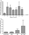
Figure 1
Figure 1
CCR3 messenger RNA (mRNA) expression on mouse bone marrow-derived mast cells (BMMC). Relative CCR3 mRNA expression on BMMC from 1–10 weeks in culture with interleukin-3 (IL-3) measured by real-time polymerase chain reaction, dashed line (––) indicates level of relative CCR3 mRNA expression on 4-week BMMC from CCR3−/− mice (a). Relative CCR3 mRNA expression on immunoglobuoin E (IgE)-activated 10-week mature c-kit+ cells (b). Data are mean ± SEM (n = 3) using RNA from three independent cultures. Statistical significance was determined using the Kruskal–Wallis test with Dunn’s multiple comparison post-test. DNP, dinitrophenol.
Figure 2
Figure 2
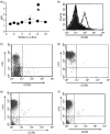
CCR3 protein expression on mouse bone marrow-derived mast cells (BMMC). Surface expression of CCR3 or isotype control on BMMC from 1–10 weeks in culture with interleukin-3 (IL-3). Data are n = 3 using cells from three independent BMMC cultures (a). Surface expression of CCR3 on mouse allergen-challenged lung cells. Closed histogram indicates isotype control and open histogram CCR3 staining (b). Intracellular expression of CCR3 on 2-week (c) and 10-week (d) BMMC. Extracellular expression of CCR3 on resting (e) and immunoglobulin E (IgE)-activated (f) 10-week mature BMMC. Representative dot plots show CCR3 expression. The upper left quadrant of the dot plots indicates the location of the c-kit phycoerythrin+, fluorescein isothiocyanate isotype control population.
Figure 3
Figure 3
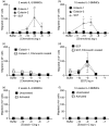
Immature and mature bone marrow-derived mast cells (BMMC) migration to eotaxin-1 (CCL11) or eotaxin-2 (CCL24). Two-week (a) and 10-week (b) BMMC were assayed for migration towards eotaxin-1 (▪) and eotaxin-2 (▴ 10 pm to 100 nm) and stem cell factor (SCF; +, 0·1–10 nm). After 3 hr incubation, migrated cells were recovered and double stained with anti-c-kit-phycoerythrin and anti-GR-1-fluorescein isothiocyanate and the c-kit+ BMMC were counted by flow cytometry. Two-week Gr-1+ cells migrated towards eotaxin-1 (▪) and eotaxin-2 (▴), 0·1 nm to 100 nm). Data are expressed as chemotactic index (number of migrated Gr-1+ cells/number of cells migrated to buffer; c) Fibronectin coating of chemotaxis membranes did not increase 2-week BMMC migration to eotaxin-1 (d) or SCF (e). FcεR1 cross-linking of BMMC did not induce migration of 2-week (f) or 10-week (g) BMMC to eotaxin-1. Data are mean ± SEM (n = 3 to n = 6) from independent BMMC cultures. *Indicates significant migration (P < 0·05) compared with buffer, **Indicates significant migration (P < 0·01) compared with buffer. Statistical significance of chemokine-induced migration compared with buffer alone was determined using Kruskal–Wallis test with Dunns post-test. Concentration response curves were compared using two-way analysis of variance with Bonferroni’s post-test.
Figure 4
Figure 4
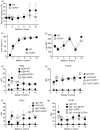
Growth and mature mast cell marker expression on CCR3−/− and wild-type bone marrow-derived mast cells (WT BMMC). WT (▪) and CCR3−/− (□) total cell number in culture over 10 weeks (a). Percentage of c-kit+ WT (▪) and CCR3−/− (□) cells over 10 weeks in culture (b). Expression of mast cell markers c-kit (c), CD34 (d), T1/ST2 (e), CD13 (f) and FcεR1 (g) were measured on WT (▪) and CCR3−/− (□) BMMC over 10 weeks in culture. Data are mean ± SEM (n = 3) from independent BMMC cultures.
Figure 5
Figure 5
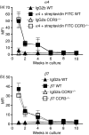
Progenitor mast cell marker expression on CCR3−/− and wild-type bone marrow-derived mast cells (WT BMMC). α4 integrin (a) and β7 (b) integrin expression on WT (▪) and CCR3−/− (□) BMMC over 10 weeks in culture. Data are mean ± SEM (n = 3) from independent BMMC cultures.
Figure 6
Figure 6
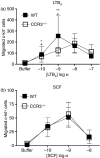
Migration of CCR3−/− and wild-type bone marrow-derived mast cells (WT BMMC) to the mast cell chemoattractants leukotriene B4 (LTB4) or stem cell factor (SCF). Chemotaxis assays of 2 week WT (▪) and CCR3−/− (□) BMMC were set up to LTB4 (a) and SCF (b). After 3 hr incubation, migrated cells were recovered and double stained with anti-c-kit-phycoerythrin and anti-GR-1-fluorescein isothiocyanate and the c-kit+ BMMCs were counted by flow cytometry. Data are mean ± SEM (n = 3) from independent BMMC cultures. *Indicates significant migration (P < 0·05), **Indicates significant migration (P < 0·01) compared with buffer for WT data, + indicates significant migration compared with buffer (P < 0·05) for knockout data. Statistical significance of chemokine-induced migration compared with buffer alone was determined using Kruskal–Wallis test with Dunns post-test. Concentration response curves were compared using two-way analysis of variance with Bonferroni’s post-test.

Similar articles

Cited by

References

    1. Sayed BA, Christy A, Quirion MR, Brown MA. The master switch: the role of mast cells in autoimmunity and tolerance. Annu Rev Immunol. 2008;26:705–39. - PubMed
    1. Fokkens WJ, Godthelp T, Holm AF, Blom H, Mulder PG, Vroom TM, Rijntjes E. Dynamics of mast cells in the nasal mucosa of patients with allergic rhinitis and non-allergic controls: a biopsy study. Clin Exp Allergy. 1992;22:701–10. - PubMed
    1. Nouri-Aria KT, Pilette C, Jacobson MR, Watanabe H, Durham SR. IL-9 and c-Kit+ mast cells in allergic rhinitis during seasonal allergen exposure: effect of immunotherapy. J Allergy Clin Immunol. 2005;116:73–9. - PubMed
    1. Brightling CE, Bradding P, Symon FA, Holgate ST, Wardlaw AJ, Pavord ID. Mast-cell infiltration of airway smooth muscle in asthma. N Engl J Med. 2002;346:1699–705. - PubMed
    1. Carroll NG, Mutavdzic S, James AL. Increased mast cells and neutrophils in submucosal mucous glands and mucus plugging in patients with asthma. Thorax. 2002;57:677–82. - PMC - PubMed

Publication types

LinkOut - more resources