. 2010 Jan 13;132(1):321-7.
doi: 10.1021/ja907966n.

Protein (19)F NMR in Escherichia coli

Affiliations

Protein (19)F NMR in Escherichia coli

Conggang Li et al. J Am Chem Soc. .

Abstract

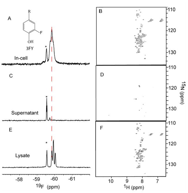
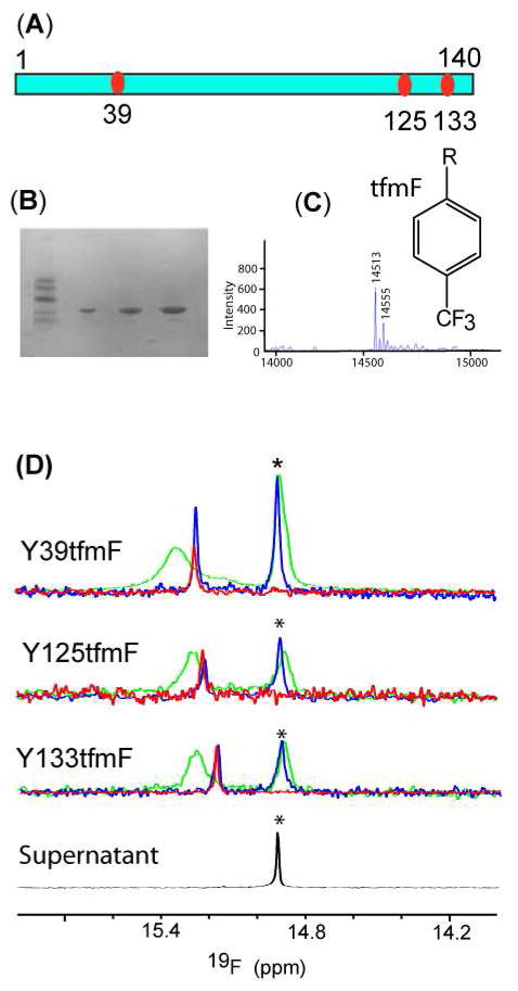
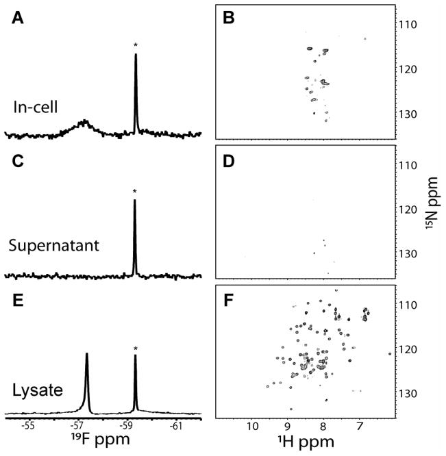
Although overexpression and (15)N enrichment facilitate the observation of resonances from disordered proteins in Escherichia coli, (15)N enrichment alone is insufficient for detecting most globular proteins. Here, we explain this dichotomy and overcome the problem while extending the capability of in-cell NMR by using (19)F-labeled proteins. Resonances from small (approximately 10 kDa) globular proteins containing the amino acid analogue 3-fluoro-tyrosine can be observed in cells, but for larger proteins the (19)F resonances are broadened beyond detection. Incorporating the amino acid analogue trifluoromethyl-L-phenylalanine allows larger proteins (up to 100 kDa) to be observed in cells. We also show that site-specific structural and dynamic information about both globular and disordered proteins can be obtained inside cells by using (19)F NMR.


Figures

Figure 1
Figure 1
19F spectra of 3FY labeled αSYN (left panels) and 1H-15N HSQC spectra of 15N-enriched αSYN (right panels). Panels A and B show in-cell spectra. The inset in panel A shows the structure of 3FY. Panels C and D show spectra of supernatants collected immediately after completing the in-cell spectra. Panels E and F show spectra of supernatants from the clear lysates. The asterisks indicate the free 3FY resonances. The dashed vertical line shows the upfield shift on cell lysis.
Figure 2
Figure 2
Sites of tfmF incorporation in α-synuclein (A). SDS-PAGE of the three purified tfmF labeled α-synucleins (B). ESI-mass spectrum of tfmF39 labeled α–synuclein (C). The inset shows the structure of tfmF. 19F spectra of labeled synuclein (D). Spectra from cell slurries are shown in green. Spectra from clear lysates are shown in blue. Spectra from purified tfmF labeled proteins are shown in red. Spectra from supernatants collected immediately after the in-cell NMR experiments are shown in black. The asterisks indicate the free tfmF resonances.
Figure 3
Figure 3
19F- and 1H-15N HSQC- spectra of 15N-enriched, 3FY labeled UBQ. The panels are labeled as described in the caption to Figure 1.
Figure 4
Figure 4
19F spectra of 3FY labeled CI2 (left panels) and 1H-15N HSQC spectra of 15N-enriched CI2. The panels are labeled as described in the caption to Figure 1.
Figure 5
Figure 5
19F spectra of K18tfmF CI2 in cells (green) and lysates (blue) (A), Y42tfmF CI2 in cells and in lysates (B), and supernatants collected after the in-cell NMR experiments (C). The asterisks indicate the free tfmF resonances.
Figure 6
Figure 6
19F- and 1H-15N HSQC- spectra of 15N-enriched, 3FY labeled CAM. The panels are labeled as described in the caption to Figure 1.
Figure 7
Figure 7
19F spectra of tfmF 39 labeled GFP in cells (green) and the purified protein in solution (blue) (A), tfmF 221 labeled GFP in cells and the purified protein in solution (B), and supernatants collected after the in-cell NMR experiments (C). The asterisks indicate the free tfmF resonances.
Figure 8
Figure 8
19F spectra of L225tfmF HDH. In-cell sample (A), purified protein (B), supernatant collected after the in-cell NMR experiments (C). The asterisks indicate the resonance from free tfmF.

Similar articles

Cited by

References

    1. Luby-Phelps K. Int Rev Cytol. 2000;192:189–221. - PubMed
    1. Malmström J, Beck M, Schmidt A, Lange V, Deutsch EW, Aebersold R. Nature. 2009;460:762–765. - PMC - PubMed
    1. Zimmerman SB, Trach SO. J Mol Biol. 1991;222:599–620. - PubMed
    1. Augustus AM, Reardon PN, Spicer LD. Proc Natl Acad Sci USA. 2009;106:5065–5069. - PMC - PubMed
    1. Brindle KM, Williams SP, Boulton M. FEBS Lett. 1989;255:121–124.

Publication types

MeSH terms