. 2009:9:1-14.
doi: 10.1673/031.009.5701.

Physiological activity of neuropeptide f on the hindgut of the blood-feeding hemipteran, Rhodnius prolixus

Affiliations

Physiological activity of neuropeptide f on the hindgut of the blood-feeding hemipteran, Rhodnius prolixus

Ronald Gonzalez et al. J Insect Sci. 2009.

Abstract

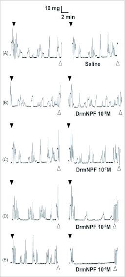
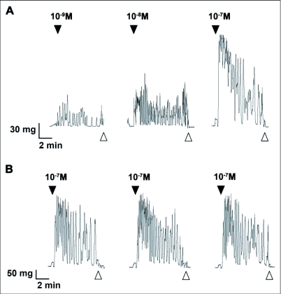
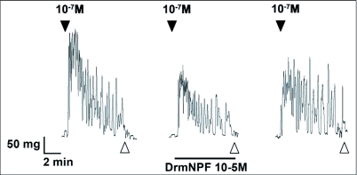
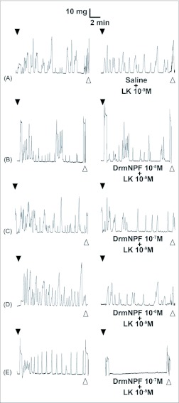
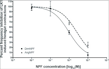
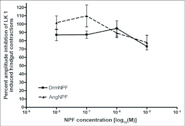
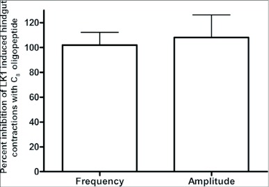
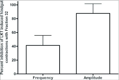
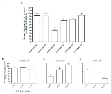
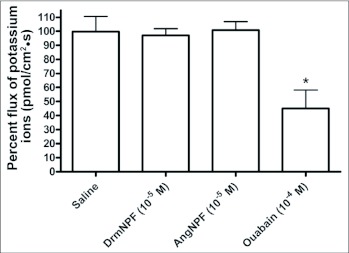
Current hypotheses propose that, in the invertebrates, neuropeptide F (NPF), the vertebrate NPY homologue, may be capable of regulating responses to diverse cues related to nutritional status and feeding. An investigation into the effects of Drosophila melanogaster NPF (DrmNPF) and Anopheles gambiae NPF (AngNPF) on hindgut physiology of Rhodnius prolixus Stal (Heimptera: Reduviidae) suggests a myoinhibitory role for these peptides and the R. prolixus native peptide. Extracts of the central nervous system of R. prolixus were processed and several HPLC-fractions revealed NPF-like activity within the nanomolar equivalent range when tested using the hindgut contraction assay. Although NPF has been shown to decrease epithelial membrane potential in Aedes aegypti larval midgut preparations, NPF does not appear to play a role in epithelial transport of potassium in the hindgut. While the function of NPF has yet to be established, NPF-like effects suggest multiple physiological roles for NPF among invertebrates.


Figures

Figure 1.
Figure 1.
Effects of increasing concentrations of DrmNPF on spontaneous contractions of a single isolated Rhodnius prolixus hindgut. Closed arrows indicate addition of saline (left column) or various concentrations of DrmNPF (right column). The addition of DrmNPF was preceded by recordings of spontaneous contractions alone (A–E). Open arrows indicate washing with physiological saline. The preparation was washed at 5 minute intervals for 20 minutes between applications of peptide solutions.
Figure 2.
Figure 2.
Leucokinin 1 (LK 1) induces contractions of Rhodnius prolixus hindgut incubated in physiological saline. (A) The effects of increasing doses of LK 1 on the phasic contractions and basal tonus of R. prolixus hindgut. At 10-9 M, LK 1 increases frequency of contractions but does not affect basal tonus of the preparation. However, increasing concentrations of LK 1 induced an increase in the frequency of phasic contractions, amplitude of contractions and a change in basal tonus. (B) Even at relatively high doses, LK 1 can induce increased myoactivity with repeated doses over time for up to 90 minutes. Closed arrows indicated addition of LK 1. Open arrows indicate washing with physiological saline. The preparations were washed at 5 minute intervals for 20 minutes between applications of peptide solutions.
Figure 3.
Figure 3.
Effects of DrmNPF at 10-5M (horizontal bar) on leucokinin 1 (LK 1)-induced contractions (10-7M) of a single isolated Rhodnius prolixus hindgut. Closed arrows indicate addition of LK 1 at 10-7 M and open arrows indicate washing with physiological saline. Preparations were washed at 5 minute intervals for 20 minutes between additions of peptide solutions. The trace shown here is a representative trace of 3 preparations.
Figure 4.
Figure 4.
Effects of increasing concentrations of DrmNPF on leucokinin 1 (LK 1)-induced contractions of a single isolated Rhodnius prolixus hindgut. Closed arrows indicate addition of 10-9 M LK 1 (left column) or various concentrations of DrmNPF and 10-9 M LK 1 (right column). The addition of DrmNPF was always preceded by a trial of LK 1 alone (A–E). Open arrows indicate washing with physiological saline. The preparation was washed at 5 minute intervals for 20 minutes between applications of peptide solutions.
Figure 5.
Figure 5.
Dose-response curve showing the effects of DrmNPF and AngNPF on the frequency of leucokinin 1 (LK 1)-induced muscle contractions of isolated Rhodnius prolixus hindgut preparations. Results are expressed as a percentage of the frequency of muscle contractions stimulated by 10-9 M LK 1 alone. The average frequency was measured by force transducer for a 4 minute interval, 1 minute after the addition of the peptide solutions. Each point represents the mean ± SEM (N = 5).
Figure 6.
Figure 6.
Dose-response curve showing the effects of DrmNPF and AngNPF on the amplitude of leucokinin 1 (LK 1)-induced muscle contractions of isolated Rhodnius prolixus hindgut preparations. Results are expressed as a percentage of the amplitude of muscle contractions stimulated by 10-9 M LK 1 alone. The average amplitude of contractions was measured for a 4 minute interval, 1 minute after the addition of the peptide solution. Each point represents the mean ± SEM (N = 5).
Figure 7.
Figure 7.
Effects of C8 oligopeptide (10-4 M) on leucokinin 1 (LK 1)-induced contractions (10-9 M) on Rhodnius prolixus hindgut. At 10-4 M, C8 had little or no effect on frequency or amplitude of contractions. Each bar represents the mean ± SEM (N =3).
Figure 8.
Figure 8.
Effects of fraction 32 on leucokinin 1 (LK 1)-induced contractions of a single isolated Rhodnius prolixus hindgut. 3.3 CNS equivalents of fraction 32 inhibited the frequency of LK 1-induced contractions and has little effect on the amplitude of hindgut contractions. Each bar represents the mean ± SEM (N = 3).
Figure 9.
Figure 9.
Effects of fractions 50–55 on leucokinin 1 (LK 1)-induced contractions of Rhodnius prolixus hindgut. (A) 3.3 CNS equivalents of fractions 50–55; fraction 52 significantly inhibited the frequency of LK 1-induced contractions. (B, C, D) Effects of increasing concentrations of fractions 50, 52, and 54 on LK 1-induced hindgut contractions. Each bar represents the mean ± SEM (N = 3, for each fraction). Statistics were performed on arcsine transformations. * p <0.05, oneway ANOVA Newman-Keuls post-test.
Figure 10.
Figure 10.
Representative example of SIET measurements showing calculated ion fluxes along the surface of the in vitro hindgut of Rhodnius prolixus. Localized ion flux was illustrated by vectors superimposed on a digital image of the hindgut. The direction of the vector reflects the movement of ions into (influx) or out of (efflux) the hindgut, whereas the length of the vector reflects the magnitude of the ion flux. Large influxes of K+ are observed over the ampulla that increased centrally and dissipated at the margins. No additional K+ fluxes were observed along the length of the hindgut. The lengths of the arrows indicate the magnitude and direction of the potassium flux (N = 10).
Figure 11.
Figure 11.
Potassium ion flux of a single isolated Rhodnius prolixus hindgut in vitro challenged with DrmNPF, AngNPF, and ouabain. The NPFs tested did not influence potassium ion transport across the ampulla of the hindgut. At 10-4 M ouabain significantly decreased potassium ion flux over the ampulla of the hindgut. Each bar represents the mean ± SEM (N = 3). Statistics were performed on arcsine transformations. * p < 0.05, one-way ANOVA Newman-Keuls post-test.

Similar articles

Cited by

References

    1. Brown MR, Raikhel AS, Lea AO. Ultrastructure of midgut endocrine cells in the adult mosquito, Aedes aegypti. Tissue and Cell. 1985;17:709–721. - PubMed
    1. Cox HM, Cuthbert AW, Håkanson R, Wahlestedt C. The effect of neuropeptide Y and peptide YY on electrogenic ion transport in rat intestinal epithelia. Journal of Physiology. 1988;398:65–80. - PMC - PubMed
    1. Donini A, O'Donnell MJ. Analysis of Na+, Cl-, K+, H+ and NH4+ concentration gradients adjacent to the surface of anal papillae of the mosquito Aedes aegypti: application of self-referencing ion-selective microelectrodes. Journal of Experimental Biology. 2005;208:603–610. - PubMed
    1. Gonzalez R, Orchard I. Characterization of neuropeptide F-like immunoreactivity in the blood-feeding hemipteran, Rhodnius prolixus. Peptides. 2008;29:545–558. - PubMed
    1. Halton DW, Shaw C, Maule AG, Smart D. Regulatory peptides in helminth parasites. Advances in Parasitology. 1994;34:163–227. - PubMed

Publication types

MeSH terms