. 2010 Mar;11(3):348-60.
doi: 10.1111/j.1600-0854.2009.01022.x. Epub 2009 Dec 3.

Gangliosides and beta1-integrin are required for caveolae and membrane domains

Affiliations

Gangliosides and beta1-integrin are required for caveolae and membrane domains

Raman Deep Singh et al. Traffic. 2010 Mar.

Abstract

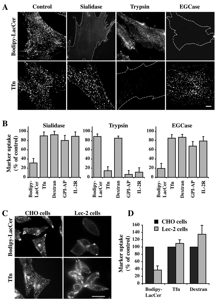
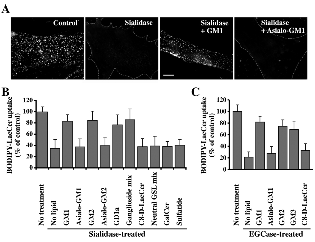
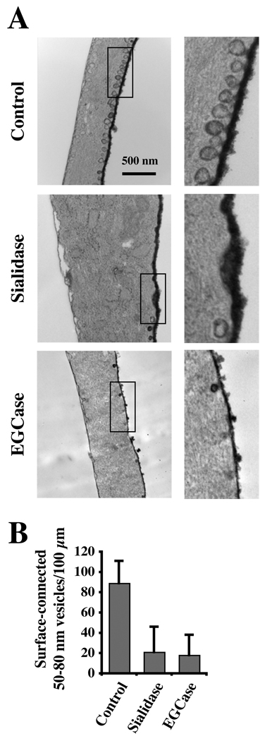
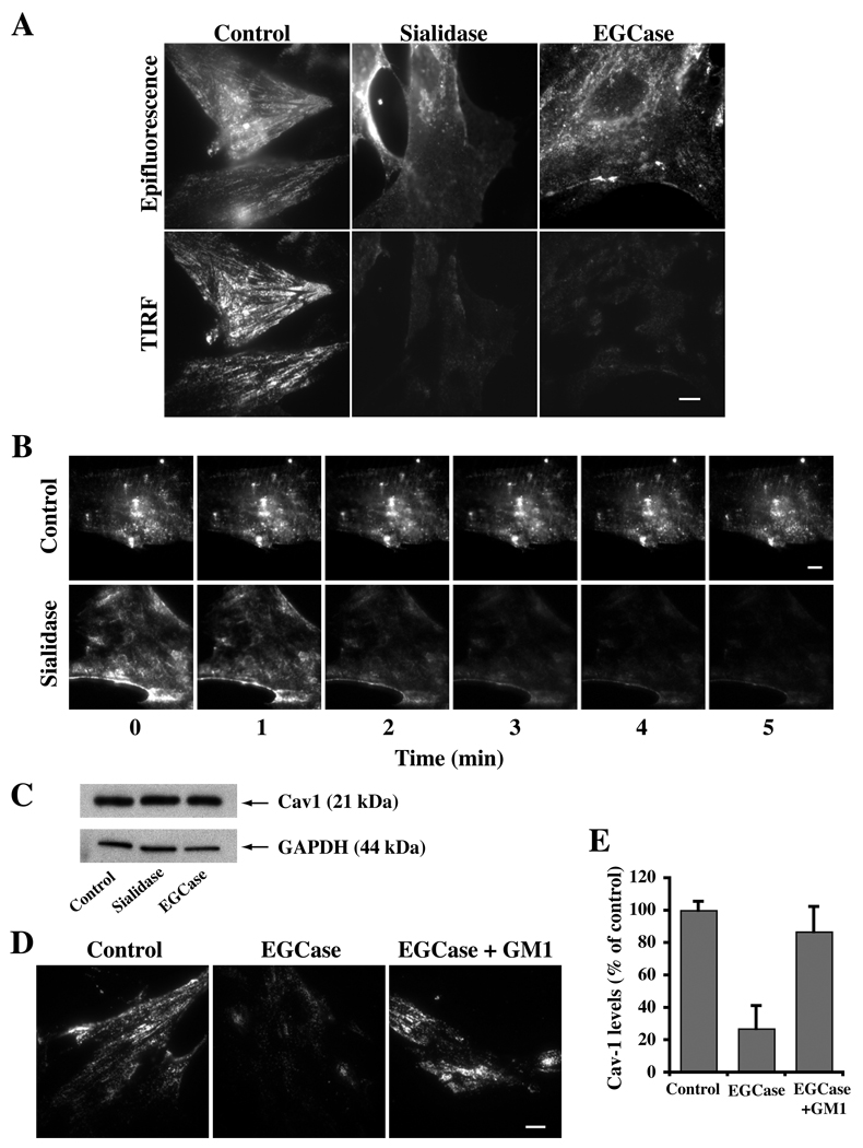
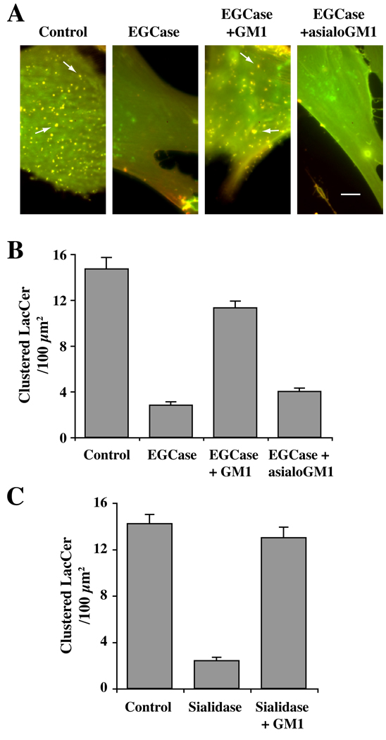
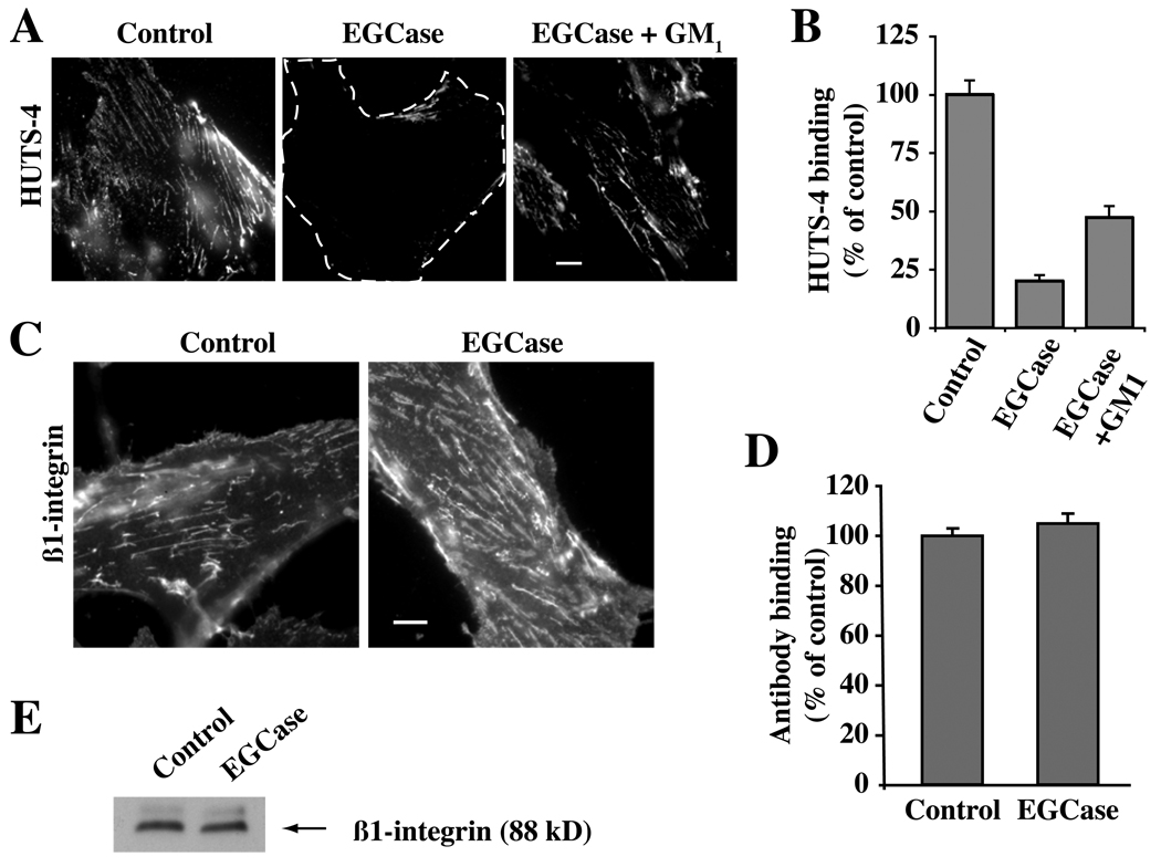
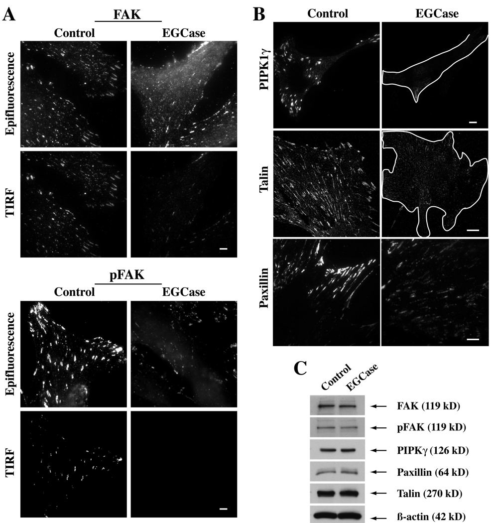
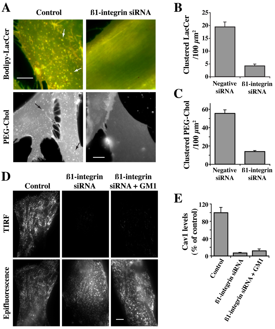
Caveolae are plasma membrane domains involved in the uptake of certain pathogens and toxins. Internalization of some cell surface integrins occurs via caveolae suggesting caveolae may play a crucial role in modulating integrin-mediated adhesion and cell migration. Here we demonstrate a critical role for gangliosides (sialo-glycosphingolipids) in regulating caveolar endocytosis in human skin fibroblasts. Pretreatment of cells with endoglycoceramidase (cleaves glycosphingolipids) or sialidase (modifies cell surface gangliosides and glycoproteins) selectively inhibited caveolar endocytosis by >70%, inhibited the formation of plasma membrane domains enriched in sphingolipids and cholesterol ('lipid rafts'), reduced caveolae and caveolin-1 at the plasma membrane by approximately 80%, and blunted activation of beta1-integrin, a protein required for caveolar endocytosis in these cells. These effects could be reversed by a brief incubation with gangliosides (but not with asialo-gangliosides or other sphingolipids) at 10 degrees C, suggesting that sialo-lipids are critical in supporting caveolar endocytosis. Endoglycoceramidase treatment also caused a redistribution of focal adhesion kinase, paxillin, talin, and PIP Kinase Igamma away from focal adhesions. The effects of sialidase or endoglycoceramidase on membrane domains and the distribution of caveolin-1 could be recapitulated by beta1-integrin knockdown. These results suggest that both gangliosides and beta1-integrin are required for maintenance of caveolae and plasma membrane domains.


Figures

Fig 1
Fig 1. Effect of sialic acid depletion on endocytosis
(A,B) HSFs were preincubated for 30 min at 37°C without (Control) or with sialidase, trypsin, or EGCase, washed, and the internalization of Bodipy-LacCer, Tfn, Dextran, GFP-GPI, and IL-2β receptor were assessed after 5 min of endocytosis at 37°C. (A) Fluorescence micrographs showing the effects of sialidase, trypsin, or EGCase pretreatments on Bodipy-LacCer and Tfn uptake. Dotted lines outline the cells in field. (B) Uptake of multiple markers after 5 min of endocytosis at 37°C. Note the selective inhibition of endocytosis of Bodipy-LacCer (but not other markers) following pretreatment with sialidase or EGCase. Endocytosis was quantified by image analysis (n ≥ 30 cells/marker in 3 independent experiments) and expressed as means ± SE relative to untreated control samples. See Fig. S2A for optimization of sialidase and EGCase pretreatment times on Bodipy-LacCer endocytosis. (C,D) Endocytosis of Bodipy-LacCer, Tfn, and dextran in a mutant CHO cell line that has decreased levels of cell surface sialic acid. (C) Fluorescence micrographs of Bodipy-LacCer and Tfn in the CHO parental (Pro5) and Lec-2 mutant cells (defective in the transport of CMP-NeuAc into the lumen of the Golgi apparatus) after 5 min of endocytosis at 37°C. (D) Quantitation of uptake by image analysis (n ≥ 30 cells/marker in 3 independent experiments). Bars, 10 µm.
Fig. 2
Fig. 2. Restoration of Bodipy-LacCer endocytosis in HSFs by sialic acid containing gangliosides
Cells were pretreated with sialidase (A,B) or EGCase (C) as in Fig. 1, washed, and pulse-labeled with Bodipy-LacCer to monitor uptake after 5 min of endocytosis at 37°C. In some instances the enzyme-pretreated cells were incubated for 30 min at 10°C with the indicated non-fluorescent lipid, washed, and subsequently pulse-labeled with Bodipy-LacCer as above. (A) Fluorescence micrographs showing inhibition of Bodipy-LacCer uptake by sialidase and restoration of endocytosis by GM1 (but not asialo-GM1) ganglioside. Bar, 10 µm. (B,C) Quantitative results for restoration of Bodipy-LacCer endocytosis by various lipids after sialidase or EGCase treatments. “No treatment,” refers to no enzymatic pretreatment; “No lipid” shows effect of enzymatic treatment without lipid addition. Values were quantified as in Fig. 1 (n ≥ 30 cells/marker in 3 independent experiments) and are expressed as means ± SE relative to untreated control samples.
Fig. 3
Fig. 3. Effect of sialidase and EGCase treatments on caveolae
(A) HSFs were untreated (Control) or treated for 30 min at 37°C with sialidase or EGCase, fixed, stained with ruthenium red to identify PM invaginations, and processed for transmission electron microscopy. Bar, 500 nm. (B) For quantitation, 50–80-nm diameter smooth, surface-connected vesicles within 0.5 µm of the cell surface were counted along the entire cell perimeter and are expressed (means ± SE) as number per 100 µm of length.
Fig. 4
Fig. 4. Cav1 is lost from the PM following sialidase or EGCase treatments but is restored by incubation with exogenous GM1 ganglioside at low temperature
(A) HSFs were untreated or treated with the indicated enzyme for 30 min at 37°C, fixed, immunostained for Cav1, and observed by epifluorescence or TIRF microscopy. Note the loss of Cav1 from the cell surface (TIRF images) in the treated cells. (B) Real time imaging of Cav1-GFP demonstrates redistribution of Cav1 from the cell surface to intracellular membranes during incubation with sialidase at 37°C. Indicated time points are taken from QuickTime movies. Control experiments using buffer alone showed no redistribution of Cav1-GFP over the same time period. (C) Total Cav1 levels by western blotting were not altered by sialidase or EGCase treatments. (D,E) Endogenous Cav1 redistributes back to the PM of EGCase-treated cells following a 30 min incubation with exogenous GM1 ganglioside at 10°C. Quantitation of TIRF signal for Cav1 in panel (E) was by image analysis (n ≥ 30 cells/condition) and is expressed (mean ± SE) as a percent of controls. Bars, 10 µm.
Fig. 5
Fig. 5. Effect of sialidase or EGCase treatment on the distribution of Cav1 and PTRF-Cavin
HSFs were treated with or without the indicated enzyme for 30 min at 37°C followed by immunostaining of Cav1 (red) and PTRF-Cavin (green). Note the typical surface-like staining of Cav1 and extensive co-localization with PTRF-Cavin in control cells, while in sialidase- or EGCase-treated cells these proteins were non-overlapping and in different structures. Arrows indicate Golgi region. Bars, 10 µm.
Fig. 6
Fig. 6. Enzymatic cleavage of cell surface sialic acid residues abolished PM domains enriched in Bodipy-LacCer
HSFs were untreated (Control), or pretreated with EGCase (A,B) or sialidase (C) for 30 min at 37°C, followed by incubation with 2.5 µM Bodipy-LacCer for 30 min at 10°C. Cells were then washed and fluorescence images were acquired simultaneously at green and red wavelengths and merged. Imaging was carried out at low temperature to inhibit endocytosis. (A) Note the presence of yellow/orange micron size clusters in control cells (e.g., at arrows) whereas no such domains were observed in EGCase-treated cells (middle panels). PM domain formation was restored when the enzyme-treated cells were incubated for 30 min at 10°C with exogenous GM1 ganglioside prior to incubation with Bodipy-LacCer (right panels). No restoration of PM domains was seen using asialo-GM1 in place of GM1. Bars, 10 µm. See Fig. S3 for an image of BODIPY-LacCer-labeled sialidase-treated cells. In (B,C) the number of “patches” ≥ 1×1 µm were quantified by image analysis and are expressed per 100 µm2 area (n ≥ 6 cells per condition in 2 or more independent experiments).
Fig. 7
Fig. 7. Effect of EGCase treatment on β1-integrin levels and activation
HSFs were grown in serum-containing medium, washed, and were either untreated (Control) or pretreated with EGCase for 30 min at 37°C. (A,B) Control or EGCase treated cells were washed, warmed for 30 sec at 37°C, fixed, and immunostained with HUTS-4 mAb to detect activated β1-integrin. In one experiment the EGCase treated cells were subsequently washed and incubated for 30 min at 10°C with GM1 ganglioside prior to HUTS-4 fixation and staining. In (B), images from the experiment in panel (A) were quantified as in panel by image analysis (n ≥ 50 cells) and are expressed as a percent of untreated control samples (mean ± SE). (C,D) Live cells were stained for 30 min at 10°C using a β1-integrin FITC-labeled antibody (monitors both activated and non-activated β1-integrin), washed, and observed under the fluorescence microscope. No significant difference in fluorescence intensity between Control and EGCase treated cells (n ≥ 50 cells/condition) was observed. (E) Cells treated as in (C,D) were lysed and blotted for β1-integrin (10 µg protein per lane). Bars, 10 µm.
Fig 8
Fig 8. Effect of EGCase treatment of HSFs on focal adhesion components
(A) Cells were grown in serum-containing medium, washed, and subsequently were untreated (Control) or pretreated with EGCase for 30 min at 37°C. The basal level of FAK and pFAK (Y397) was then assessed by antibody staining using both epifluorescence and TIRF microscopy. (B) Parallel cell samples, grown under the same conditions, were immunostained for talin or paxilin. In the case of PIPK1γ cells were transfected overnight with HA-tagged PIPKIγ prior to EGCase treatment, fixation and immunostaining. PIPKIγ and talin images were by confocal microscopy; paxilin images were by TIRF microscopy. (C) In parallel dishes, cells were untreated (Control) or pretreated with EGCase for 30 min at 37°C, lysed, and cell lysates (10 µg protein per lane) were immunoblotted for various proteins as indicated. Bars, 10 µm.
Fig. 9
Fig. 9. Effect of β1-integrin knockdown on PM domains and the distribution of Caveolin-1
HSFs were transfected for 72 hrs with negative (Control) or β1-integrin siRNA (Supp. Fig. S4). (A–C) Cells were then incubated with Bodipy-LacCer or AF647-PEG-Chol to visualize PM clusters (e.g., at arrows of Control samples). In (B,C) the number of micron size clusters of Bodipy-LacCer or PEG-Chol were quantified as in Fig. 6. (D,E) Cells as above were fixed and immunostained for Cav1. Samples were then visualized by TIRF or epifluorescence microscopy. In (E) TIRF images from the experiment in panel (D) were quantified by image analysis (n ≥ 50 cells) and are expressed as a percent of untreated control samples (mean ± SE). Bars, 10 µm

Similar articles

Cited by

References

    1. Mayor S, Pagano RE. Pathways of clathrin-independent endocytosis. Nat Rev Mol Cell Biol. 2007;8:603–612. - PMC - PubMed
    1. Doherty GJ, McMahon HT. Mechanisms of endocytosis. Annu Rev Biochem. 2009;78:857–902. - PubMed
    1. Hansen CG, Nichols BJ. Molecular mechanisms of clathrin-independent endocytosis. J Cell Sci. 2009;122(Pt 11):1713–1721. - PMC - PubMed
    1. Pelkmans L, Helenius A. Endocytosis via caveolae. Traffic. 2002;3:311–320. - PubMed
    1. Hill MM, Bastiani M, Luetterforst R, Kirkham M, Kirkham A, Nixon SJ, Walser P, Abankwa D, Oorschot VM, Martin S, Hancock JF, Parton RG. PTRF-Cavin, a conserved cytoplasmic protein required for caveola formation and function. Cell. 2008;132(1):113–124. - PMC - PubMed

Publication types

LinkOut - more resources