. 2010 Jan 5:8:1.
doi: 10.1186/1741-7007-8-1.

THOC5/FMIP, an mRNA export TREX complex protein, is essential for hematopoietic primitive cell survival in vivo

Affiliations

THOC5/FMIP, an mRNA export TREX complex protein, is essential for hematopoietic primitive cell survival in vivo

Annalisa Mancini et al. BMC Biol. .

Abstract

Background: The transcription/export complex is evolutionarily conserved from yeast to man and is required for coupled transcription elongation and nuclear export of mRNAs. FMIP(Fms interacting protein) is a member of the THO (suppressors of the transcriptional defects of hpr1delta by overexpression) complex which is a subcomplex of the transcription/export complex. THO complex (THOC) components are not essential for bulk poly (A)+ RNA export in higher eukaryotes, but for the nuclear export of subset of mRNAs, however, their exact role is still unclear.

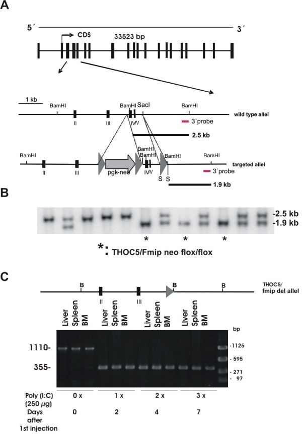
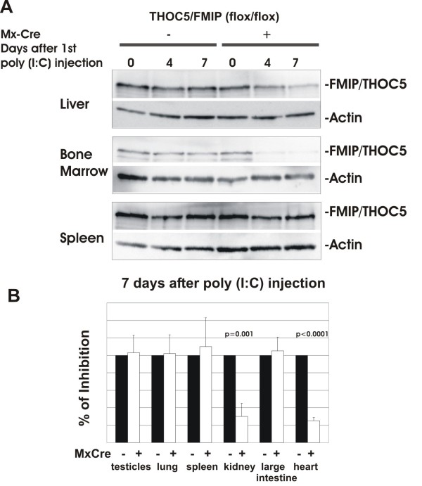
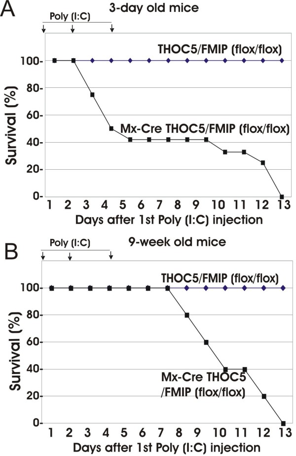
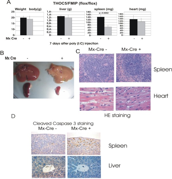
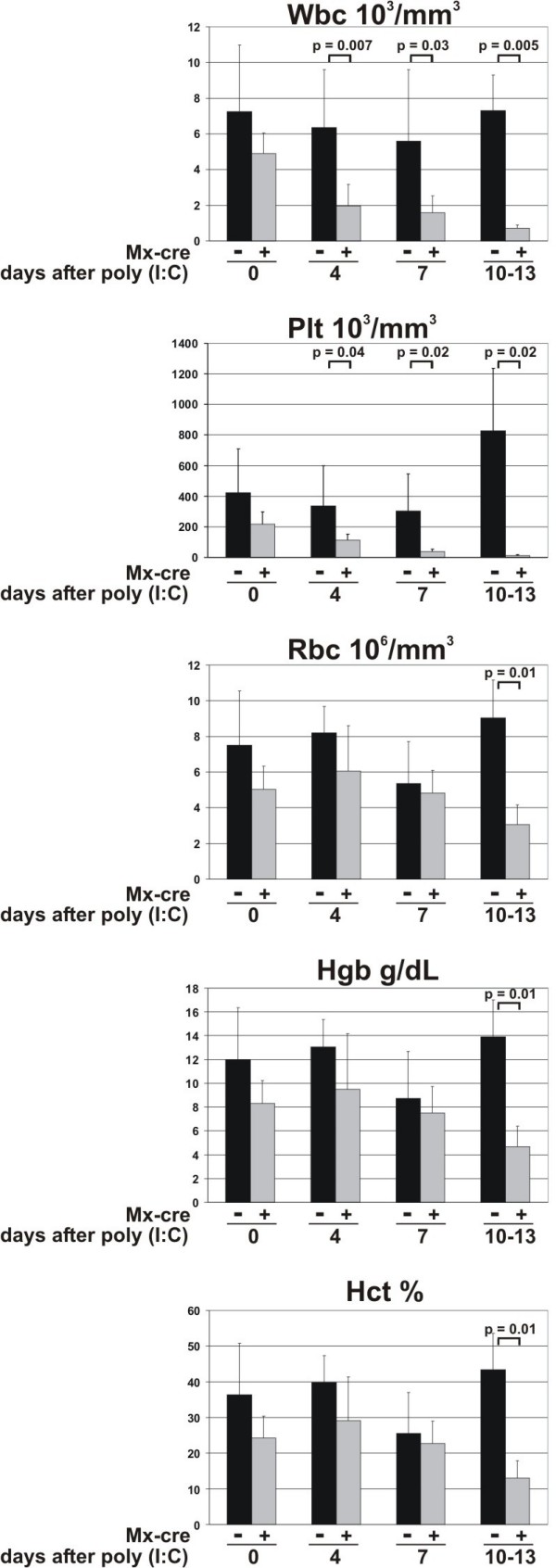
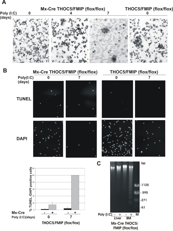
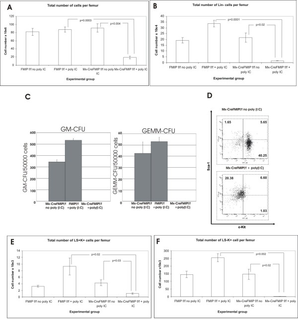
Results: To study the role of THOC5/Fms interacting protein in vivo, we generated THOC5/Fms interacting protein knockout mice. Since these mice are embryonic lethal, we then generated interferon inducible conditional THOC5/Fms interacting protein knockout mice. After three poly injections all of the mice died within 14 days. No pathological alterations, however, were observed in liver, kidney or heart. Thus we considered the hematopoietic system and found that seven days after poly injection, the number of blood cells in peripheral blood decreased drastically. Investigation of bone marrow cells showed that these became apoptotic within seven days after poly injection. Committed myeloid progenitor cells and cells with long term reconstituting potential were lost from bone marrow within four days after poly injection. Furthermore, infusion of normal bone marrow cells rescued mice from death induced by loss of THOC5/Fms interacting protein.

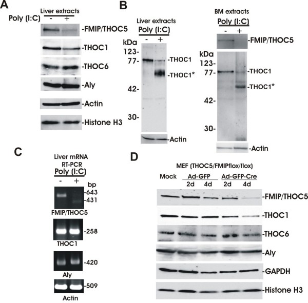
Conclusion: THOC5/Fms interacting protein is an essential element in the maintenance of hematopoiesis. Furthermore, mechanistically depletion of THOC5/Fms interacting protein causes the down-regulation of its direct interacting partner, THOC1 which may contribute to altered THO complex function and cell death.


Figures

Figure 1
Figure 1
Generation of THOC5/FMIP (flox/flox) and Mx-cre THOC5/FMIP (flox/flox) mice. (A) The genomic structure of the 33,523 kb THOC5/FMIP locus is depicted. THOC5/FMIP deficient mice were generated using a genomic DNA fragment containing exons II, III, IV, and V isolated from 129/ola genetic background. The targeting vector was constructed from a 3.9 kb PCR fragment and an adjacent 2.2 kb fragment harboring exons IV/V. Fragments were introduced into the pPNTloxPneo vector via NotI, XhoI restriction sites and KpnI, respectively. An additional loxP site was inserted into the SacI site downstream of exon V: (B) After transfection, ES cell clones carrying the inserted floxed neo cassette and floxed Fmip exons IV/V were identified by the presence of an 1.9 kb BamHI restriction fragment in Southern Blot analysis with external 3'probe ((*) THOC5/fmip neo flox/flox). (C) Mx-cre THOC5/FMIP (flox/flox) were sacrificed before (zero day) and after two (one × poly (I:C) injection), four (two × poly (I:C) injection) and seven (three × poly (I:C) injection) days after the first poly (I:C) injection and genomic DNAs were extracted from liver, spleen, and bone marrow. Genomic DNAs were supplied for the deletion of exons IV/V determined by PCR using 5'-TGCTGGCATTGAACTGTG-3' and 5'-CAGCACTGGAGCGGGAGATGT-3'. PCR product: wild type allel: 1110 bp; THOC5/FMIP del allele: 355 bp.
Figure 2
Figure 2
THOC5/FMIP expression is reduced in specific organs. (A, B) 250 μg of poly (I:C) were injected into six-week-old Mx-cre THOC5/FMIP (flox/flox) (n = 8) or control THOC5/FMIP (flox/flox) (n = 6) mice three times at two-to-three-day intervals. THOC5/FMIP protein levels in liver, bone marrow, spleen (A), testicles, lung, kidney, large intestine, and heart (B) were examined by THOC5/FMIP specific immunoblotting. Proteins were extracted from each organ. Total protein amount was standardized by actin specific immunoblotting.
Figure 3
Figure 3
Deletion of THOC5/FMIP is lethal in adult mice. Three day- (n = 25) (A) and nine-week-old (n = 7) (B) mice were injected with 50, and 500 μg of poly (I:C), respectively. Injection was performed i.p. three times at two to three day intervals. (arrows: poly (I:C) injection).
Figure 4
Figure 4
THOC5/FMIP deletion causes reduction of spleen size and induction of cleaved caspase 3. Two hundred and fifty micrograms of poly (I:C) were injected into six-week-old Mx-cre THOC5/FMIP (flox/flox) (n = 9) or control THOC5/FMIP (flox/flox) (n = 6) mice three times at two-to-three-day intervals. (A) Weight of body, liver, spleen and heart. (B) Macrography of liver and spleen: seven days after the first poly (I:C) treatment. Mx-cre -: control mice (THOC5/FMIP (flox/flox)); Mx-cre +: THOC5/FMIP depleted mice (Mx-cre THOC5/FMIP (flox/flox)). (C, D) Seven days after the first poly (I:C) injection (3×), mouse spleens, heart, liver were fixed in formalin. Paraffin sections were stained by hematoxylin and eosin (C). Spleen and liver sections were supplied for immunohitochemical staining using cleaved caspase 3 (D). Original magnification: ×100 for C and ×200 for D.
Figure 5
Figure 5
The effect of deletion of THOC5/FMIP on hematopoiesis. Six-week-old Mx-cre THOC5/FMIP (flox/flox) mice (Mx cre +, n = 9) and THOC5/FMIP (flox/flox) mice (Mx-cre -, n = 6) were injected with poly (I:C) (250 μg each) and blood was taken from the tail vein on zero, four and seven days after the first poly (I:C) injection. In addition, 10-13 days after poly (I:C) injection blood samples from Mx-cre THOC5/FMIP (flox/flox) mice (n = 3) which show severe symptom and control THOC5/FMIP (flox/flox) (n = 3) mice were taken from tails. Samples were analyzed on an ABC Vet automated blood counter. Wbc = white blood cells (103/mm3); Plt = platelets (103/mm3); Rbc = red blood cells (106/mm3); Hgb = hemoglobin (g/dl); Hct = hematocrit (%).
Figure 6
Figure 6
Deletion of THOC5/FMIP causes apoptosis of leukocytes in bone marrow. Six-week-old Mx-cre THOC5/FMIP (flox/flox) mice (n = 9) and THOC5/FMIP (flox/flox) control mice (n = 6) were injected with poly (I:C) (250 μg each) and the femora were then isolated. (A): Bone marrow cells were spun down onto glass slides and then stained with May-Grunwald Giemsa and hematoxylin. (B): Aliquots of same preparation were stained with TUNEL and DAPI. Results are the mean +/- SEM of %TUNEL positive/DAPI positive cells (n >2000 cells). Original magnification: ×200 for all panels. (C): Aliquots of 2-3 μg of DNA from liver and bone marrow of poly (I:C) treated (+) and untreated (-) Mx-cre THOC5/FMIP (flox/flox) mice were separated on 1.5% (w/v) agarose gel, stained with ethidium bromide (2 μg/ml) and photographed under UV light. M: base pair Marker.
Figure 7
Figure 7
Deletion of THOC5/FMIP causes loss of primitive hematopoietic cells from the bone marrow. Five-to-six-week-old Mx-cre THOC5/FMIP (flox/flox) (n = 5) and THOC5/FMIP (flox/flox) (n = 6) mice were injected with poly (I:C) (2 × 500 μg, two days interval). Then, bone marrow cells were isolated from mice four days after poly (I:C) injection. The effect of THOC5/FMIP depletion on the total numbers of primitive cells present in the bone marrow was assessed using a number of different approaches (A; B): total bone marrow cellularity per femur was assessed (A) and the number of cells present in the bone marrow which do not express lineage markers (B) (and therefore have a primitive phenotype) was calculated (Mean+/-SEM). Results shown are for Mx-cre THOC5/FMIP (flox/flox) (Mx-CreFMIPf/f) mice with (n = 5) and without (n = 4) poly (I:C) (2 × 500 μg, two day interval), and THOC5/FMIP (flox/flox) (FMIPf/f) control mice with (n = 6) and without (n = 6) Poly (I:C) (2 × 500 μg, two day interval) (C). The number of colony forming cells per femur was determined for Colony Forming Unit-Granulocyte macrophage (GM-CFU) and Colony Forming Unit-Granulocyte Erythroid Macrophage Megakaryocyte (GEMM-CFU). Results shown are the mean+/-SEM, n = 3. (D): Flow cytometric profile of Kit and Sca staining in Lineage marker depleted cells from mice treated with and without poly (I:C) (3 × 250 μg, two day interval). The results shown are representative of six experiments. (E; F) Results shown are for Mx-cre THOC5/FMIP (flox/flox) mice with (n = 5) and without (n = 4) poly (I:C) (2 × 500 μg, two day interval), and THOC5/FMIP (flox/flox) control mice with (n = 6) and without (n = 6) poly (I:C) (2 × 500 μg, two-day-interval), and are expressed as total number of LSK cells (E) and Lin-Sca-Kit+ cells (LS-K+) cells (F) per femur, error bars show SEM.
Figure 8
Figure 8
Bone marrow transplantation rescues THOC5/FMIP depleted mice from death. (A) Bone marrow (BM) transplantation: 106 bone marrow cells obtained from THOC5/FMIP (flox/flox) mice (n = 14) were transferred into six-to-eight-week-old nonablated Mx-cre THOC5/FMIP (flox/flox) mouse for each and one day after transplantation recipient mice were injected with 250 μg of poly (I:C). Injection was performed i.p. three times at two-to-three-day intervals. No transplantation: Mx-cre THOC5/FMIP (flox/flox) mice without BM transplantation were injected with poly (I:C). (B) Bone marrow cells were spun down onto glass slides and then stained with May-Grunwald Giemsa and hematoxylin. Bone marrow cells were derived from 1) non-treated Mx-cre THOC5/FMIP (flox/flox) mouse. 2) a poly (I:C) injected normal bone marrow cell recipient Mx-cre THOC5/FMIP (flox/flox) mouse which died 18 days after the first injection (Nonsurvivor). 3) a poly (I:C) injected normal bone marrow cell recipient Mx-cre THOC5/FMIP (flox/flox) survivor (eight weeks after the first injection) (Survivor).
Figure 9
Figure 9
Depletion of THOC5/FMIP causes down-regulation of THOC1. (A): Liver extracts from three times poly (I:C) 250 μg treated (+) (14 days) and untreated (-) six-week-old Mx-cre THOC5/FMIP (flox/flox) mice (n = 3) were examined by THOC1, Aly, THOC6, THOC5/FMIP, Actin and Histone H3 specific immunoblotting. Each sample consisted of 100 μg of liver. (B): Liver or bone marrow extracts from poly (I:C) treated (+) (14 days) were examined by THOC1, THOC5/FMIP, and actin specific immunoblotting. THOC1*: 60 kDa THOC1 degradation product from poly (I:C) treated liver or bone marrow sample. (C): mRNA was isolated from the same liver samples and THOC5/FMIP (643 bp and 431 bp (without exons IV/V)), THOC1 (258 bp), Aly (420 bp) and Actin (509 bp) specific RT-PCR were performed. (D): MEF THOC5/FMIP (flox/flox) cells were infected with adenovirus carrying GFP gene (Ad-GFP) or adenovirus carrying GFP and cre-recombinase genes (Ad-GFP-Cre) (M.O.I. 50; Vector Biolab, Philadelphia, PA, USA) then lysed by laemmli buffer. Extracts were then examined by THOC1, Aly, THOC6, THOC5/FMIP, GAPDH and Histone H3 specific immunoblotting.

Similar articles

Cited by

References

    1. Reed R, Hurt E. A conserved mRNA export machinery coupled to pre-mRNA splicing. Cell. 2002;108:523–531. doi: 10.1016/S0092-8674(02)00627-X. - DOI - PubMed
    1. Aguilera A. Cotranscriptional mRNP assembly: from the DNA to the nuclear pore. Curr Opin Cell Biol. 2005;17:242–250. doi: 10.1016/j.ceb.2005.03.001. - DOI - PubMed
    1. Reed R, Cheng H. TREX, SR proteins and export of mRNA. Curr Opin Cell Biol. 2005;17:269–273. doi: 10.1016/j.ceb.2005.04.011. - DOI - PubMed
    1. Kohler A, Hurt E. Exporting RNA from the nucleus to the cytopasm. Nat Rev Mol Cell Biol. 2007;8:761–773. doi: 10.1038/nrm2255. - DOI - PubMed
    1. Rehwinkel J, Herold A, Gari K, Kocher T, Rode M, Ciccarelli FL, Wilm M, Izaurralde E. Genome-wide analysis of mRNAs regulated by the THO complex in Drosophila melanogaster. Nat Struct Mol Biol. 2004;6:558–566. doi: 10.1038/nsmb759. - DOI - PubMed

Publication types

MeSH terms