. 2010 Jun;48(6):1169-79.
doi: 10.1016/j.yjmcc.2009.12.019. Epub 2010 Jan 4.

KChIP2 attenuates cardiac hypertrophy through regulation of Ito and intracellular calcium signaling

Affiliations

KChIP2 attenuates cardiac hypertrophy through regulation of Ito and intracellular calcium signaling

Hongwei Jin et al. J Mol Cell Cardiol. 2010 Jun.

Abstract

Recent evidence shows that the auxiliary subunit KChIP2, which assembles with pore-forming Kv4-subunits, represents a new potential regulator of the cardiac calcium-independent transient outward potassium current (I(to)) density. In hypertrophy and heart failure, KChIP2 expression has been found to be significantly decreased. Our aim was to examine the role of KChIP2 in cardiac hypertrophy and the effect of restoring its expression on electrical remodeling and cardiac mechanical function using a combination of molecular, biochemical and gene targeting approaches. KChIP2 overexpression through gene transfer of Ad.KChIP2 in neonatal cardiomyocytes resulted in a significant increase in I(to)-channel forming Kv4.2 and Kv4.3 protein levels. In vivo gene transfer of KChIP2 in aortic banded adult rats showed that, compared to sham-operated or Ad.beta-gal-transduced hearts, KChIP2 significantly attenuated the developed left ventricular hypertrophy, robustly increased I(to) densities, shortened action potential duration, and significantly altered myocyte mechanics by shortening contraction amplitudes and maximal rates of contraction and relaxation velocities and decreasing Ca(2+) transients. Interestingly, blocking I(to) with 4-aminopyridine in KChIP2-overexpressing adult cardiomyocytes significantly increased the Ca(2+) transients to control levels. One-day-old rat pups intracardially transduced with KChIP2 for two months then subjected to aortic banding for 6-8 weeks (to induce hypertrophy) showed similar echocardiographic, electrical and mechanical remodeling parameters. In addition, in cultured adult cardiomyocytes, KChIP2 overexpression increased the expression of Ca(2+)-ATPase (SERCA2a) and sodium calcium exchanger but had no effect on ryanodine receptor 2 or phospholamban expression. In neonatal myocytes, KChIP2 notably reversed Ang II-induced hypertrophic changes in protein synthesis and MAP-kinase activation. It also significantly decreased calcineurin expression, NFATc1 expression and nuclear translocation and its downstream target, MCiP1.4. Altogether, these data show that KChIP2 can attenuate cardiac hypertrophy possibly through modulation of intracellular calcium concentration and calcineurin/NFAT pathway.


Figures

Figure 1
Figure 1. KChIP2 increases Kv4.2 and Kv4.3 expression
Representative immunoblots of individual experiments showing neonatal rat myocytes express far less KChIP2 compared to adult myocytes(A); the expression of Kv4.2 and Kv4.3 is increased following KChIP2 overexpression in neonatal myocytes compared to β-gal (B). Kv4.3 induction shows two bands (66 and ~45/50 kDa). Protein expression was normalized to GAPDH. Bar graphs of fold change in the protein levels are shown in (C). (D) Representative RT-PCR and (E) quantification of mRNA expression of Kv4.2, Kv4.3 and Kv1.4 normalized to GAPDH mRNA (n=4). Data from 3–4 experiments.#, * P< 0.05 Ad.KChIP2 vs. Ad.β-gal.
Figure 2
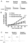
Figure 2. In vivo acute KChIP2 overexpression reverses hypertrophy-induced electrical remodeling
Representative whole-cell outward currents (A) and average current-voltage relationships of Ito (B) recorded from sham or banded hearts infected with β-gal or KChIP2 for 8 days. Data points are means±SEM; Sham vs. Ad.KChIP2, P<0.05; Ad.KChIP2 vs. Ad.β-gal, P<00.1. (C) Representative AP traces from sham or banded hearts infected with β-gal or KChIP2. Zero current potential of single myocytes ranged between −76 to 83mV. There are no significant different among the sham (−79.1+8.4 mV, n=20) β-gal groups (−76.1+6.2mV, n=18) and KChIP2 groups (−77.8+ 4.8 mV, n=18). (D) Representative immunoblots of KChIP2, Kv4.2 and Kv4.3 expression in the above hearts with summarized quantitative data (±SEM) of the relative protein density in arbitrary units (E). GAPDH levels are shown to verify protein loading and used to normalize the expression levels.
Figure 3
Figure 3. In vivo KChIP2 expression alters cardiomyocyte mechanics
KChIP2 effects on cardiomyocyte contractility and Ca2+ transients were determined in cardiomyocytes isolated from banded hearts infected with β-gal or KChIP2 in the presence of 1–10 mM Ca2+ compared to sham hearts., overexpression of KChIP2 significantly decreased cell shortening (A, n=25), velocities of contraction (B, n=25) and relaxation (C, n=25) and induced shorter Ca2+ amplitudes (D, n=21) compared to β-gal (data shown at 4 mM Ca2+). * P< 0.01 Ad.β-gal vs. Sham; § P<0.04, Ad.KChIP2 vs. Ad.β-gal.
Figure 4
Figure 4. KChIP2 regulation of calcium cycling proteins and intracellular Ca2+
The effect of KChIP2 on the expression of major Ca2+ cycling proteins were examined by immunoblot analysis in isolated adult cardiomyocytes infected with Ad.β-gal or Ad.KChIP2. (A) Western blots of myocyte lysates demonstrating expression of KChIP2, Serca2a, phospholamban (PLB), Na+-Ca2+ exchanger (NCX), and ryanodine receptor 2 (RyR2). GAPDH, an internal loading control, is used to normalize protein expression. (B) Densitometric analysis of mean data (± SEM). (C) Histogram comparing mean (±SEM) Ca2+ ratio determined between β-gal- and KChIP2-infected cardiomyocytes in the absence or presence of 4 mM 4-aminopyridine (4-AP). Blocking Ito with 4-AP in KChIP2-overexpressing adult cardiomyocytes significantly increased the Ca2+ transients. * P< 0.05 Ad.KChIP2 vs. Ad.β-gal; # P< 0.0001 Ad.KChIP2 vs. Ad.β-gal, n=12; § P< 0.001 Ad.KChIP2 vs. Ad.KChIP2+4-AP, n=15.
Figure 5
Figure 5. KChIP2 inhibits protein synthesis
The effects of KChIP2 on protein synthesis were evaluated by measuring the rate of 3H-Leucine incorporation in neonatal myocytes uninfected (control) or infected with Ad.β-gal or Ad.KChIP2 in the absence (−) or presence (+) of Ang II stimulation (1 µM). KChIP2 overexpression significantly blocked the pronounced increase in Ang II-induced 3H-Leucine incorporation. Data from five individual experiments are shown. * P< 0.001 Cont+AngII vs. Cont; #P<0.01 Ad.β-gal+Ang II vs. Ad.β-gal; §P<0.01 Ad.KChIP2+AngII vs. Ad.β-gal+AngII.
Figure 6
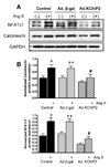
Figure 6. KChIP2 inhibits calcineurin/NFAT pathway
The effect of KChIP2 on NFATc1 and calcineurin expression (A) was evaluated by Western blotting in neonatal myocytes infected with Ad.β-gal or Ad.KChIP2 (MOI 60) in the presence (+) or absence (−) of Ang II. (B) Quantitative analysis of mean data (± SEM) of 3 to 4 independent experiments. GAPDH, an internal loading control, is used to normalize protein expression. * P< 0.05 Cont+AngII vs. Cont; **P<0.01 Ad.β-gal+Ang II vs. Ad.β-gal; #P<0.01 Ad.KChIP2 vs. Ad.β-gal. (C) Control NRVM non-infected or NRVM infected with Ad.β-gal, Ad.VIVIT (a calcineurin/NFAT-specific inhibitor) or Ad.KChIP2 were stimulated with Ang II (+) for 30 min and analyzed for NFATc1 translocation. Myocyte nuclei are visualized with DAPI staining. (D) NFAT-luciferase activity was measured in NRVM infected with Ad.β-gal, Ad.KChIP2 or Ad.VIVIT in the absence (−) or presence (+) of Ang II (1 µM for 24 hours). Data were normalized to unstimulated β-gal (−AngII). *P=0.021 Ad.KChIP2+AngII vs. Ad.β-gal+AngII; #P=0.014 Ad.VIVIT+AngII vs. Ad.β-gal+AngII. (E) MCiP1.4 mRNA expression was determined by real time-PCR and normalized against GAPDH mRNA in control NRVM and NRVM infected with Ad.β-gal or Ad.KChIP2 and cultured in serum-free medium in the absence (−) or presence (+) of Ang II (1 µM for 24 hours). Density values of corresponding bands from at least six-eight independent experiments were normalized to control unstimulated (−AngII) and are expressed as mean ± SEM. * P< 0.05 Cont+AngII vs. Cont; *P<0.01 Ad.β-gal+AngII vs. Ad.β-gal; #P<0.01 Ad.KChIP2+AngII vs. Ad.β-gal+AngII; P<0.01 Ad.KChIP2+AngII vs. Cont+AngII.
Figure 7
Figure 7. KChIP2 inhibits MAPkinases activity
The effect of KChIP2 on ERK (A) and JNK (B) activities was evaluated in neonatal myocytes infected with Ad.β-gal or Ad.KChIP2 in the presence (+) or absence (−) of AngII (1 µM for 30 min). Immunoblots were probed with phospho-specific antibodies against ERK and JNK and then re-probed for the corresponding total protein that confirmed equivalent protein loadings. Shown are representative blots from 4 determinations. * P< 0.01 Ad.KChIP2 vs. Ad.β-gal.

Similar articles

Cited by

References

    1. Nabauer M, Kaab S. Potassium channel down-regulation in heart failure. Cardiovascular research. 1998 Feb;37(2):324–334. - PubMed
    1. Tomaselli GF, Marban E. Electrophysiological remodeling in hypertrophy and heart failure. Cardiovascular research. 1999 May;42(2):270–283. - PubMed
    1. Wickenden AD, Kaprielian R, Kassiri Z, Tsoporis JN, Tsushima R, Fishman GI, et al. The role of action potential prolongation and altered intracellular calcium handling in the pathogenesis of heart failure. Cardiovascular research. 1998 Feb;37(2):312–323. - PubMed
    1. Gidh-Jain M, Huang B, Jain P, el-Sherif N. Differential expression of voltage-gated K+ channel genes in left ventricular remodeled myocardium after experimental myocardial infarction. Circulation research. 1996 Oct;79(4):669–675. - PubMed
    1. Kaprielian R, Wickenden AD, Kassiri Z, Parker TG, Liu PP, Backx PH. Relationship between K+ channel down-regulation and [Ca2+]i in rat ventricular myocytes following myocardial infarction. The Journal of physiology. 1999 May 15;517(Pt 1):229–245. - PMC - PubMed

Publication types