. 2010 Feb;16(2):382-93.
doi: 10.1261/rna.1955410. Epub 2010 Jan 5.

Enzymatic and nonenzymatic functions of viral RNA-dependent RNA polymerases within oligomeric arrays

Affiliations

Enzymatic and nonenzymatic functions of viral RNA-dependent RNA polymerases within oligomeric arrays

Jeannie F Spagnolo et al. RNA. 2010 Feb.

Abstract

Few antivirals are effective against positive-strand RNA viruses, primarily because the high error rate during replication of these viruses leads to the rapid development of drug resistance. One of the favored current targets for the development of antiviral compounds is the active site of viral RNA-dependent RNA polymerases. However, like many subcellular processes, replication of the genomes of all positive-strand RNA viruses occurs in highly oligomeric complexes on the cytosolic surfaces of the intracellular membranes of infected host cells. In this study, catalytically inactive polymerases were shown to participate productively in functional oligomer formation and catalysis, as assayed by RNA template elongation. Direct protein transduction to introduce either active or inactive polymerases into cells infected with mutant virus confirmed the structural role for polymerase molecules during infection. Therefore, we suggest that targeting the active sites of polymerase molecules is not likely to be the best antiviral strategy, as inactivated polymerases do not inhibit replication of other viruses in the same cell and can, in fact, be useful in RNA replication complexes. On the other hand, polymerases that could not participate in functional RNA replication complexes were those that contained mutations in the amino terminus, leading to altered contacts in the folded polymerase and mutations in a known polymerase-polymerase interaction in the two-dimensional protein lattice. Thus, the functional nature of multimeric arrays of RNA-dependent RNA polymerase supplies a novel target for antiviral compounds and provides a new appreciation for enzymatic catalysis on membranous surfaces within cells.


Figures

FIGURE 1.
FIGURE 1.
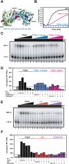
Oligomerization properties of poliovirus 3D polymerase in solution and effects of titration of wild-type and YGAA mutant polymerases into subthreshold concentrations of wild-type polymerase. (A) A ribbon diagram of the three-dimensional structure of the 461-amino acid poliovirus RNA-dependent RNA polymerase is shown with the canonical “thumb,” “fingers,” and “palm” polymerase domains indicated. Asp328 and Asp329 at the active site are shown in yellow; both of these residues were mutated to Ala residues in the YGAA mutant polymerase. Residues Leu446, Arg455, and Arg456, involved in the “thumb” surface contact of a polymerase–polymerase interaction termed Interface I are shown in blue. Residues Asp339, Ser341, and Asp349, involved in the “palm” surface of Interface I are shown in pink; these are difficult to see in this view (see also Fig. 3A). (B) Concentrated wild-type poliovirus polymerase stored in a solution that contained 50% glycerol and 140 mM NaCl was diluted to the concentrations indicated in a solution containing 15% glycerol and 40 mM NaCl. Absorbance measurements were taken every 30 sec over a 10-min time period at 350 nm. (C) Concentrated wild-type and YGAA mutant poliovirus polymerase were diluted to concentrations of 5.6 μM and turbidity was monitored. (D) Electrophoretic mobility of HP1 RNA, a 110-nt RNA derived from the 3′ end of the poliovirus genome with several U residues at its 3′ end for self-priming, is shown following incubation for 30 min with increasing amounts of YGAA mutant polymerase (lanes 2–9, containing 70, 130, 250, 500, 750, 1000, 1500, or 1750 nM YGAA mutant polymerase, respectively). (E) To a preparation of 32P-labeled HP1 RNA (lane 1), various concentrations and mixtures of wild-type and YGAA mutant poliovirus polymerase were added, incubated for 30 min, and the products were displayed (lanes 2–13). Each of lanes 2–13 contained 7.5 nM wild-type polymerase, a concentration too low to support detectable template utilization under these conditions (lanes 2,8). This was supplemented with increasing amounts of wild-type 3D polymerase (lanes 3–7, containing an additional 7.5, 22.5, 55, 117.5, or 242.5 nM wild-type polymerase, respectively) or YGAA mutant polymerase (lanes 9–13) at the same concentrations. (F) The percentage of template elongation is shown as a function of the ratio of added:basal polymerase for both added wild-type and added YGAA mutant polymerase. “WT + WT” and “YGAA + YGAA” show the activities when these polymerases were present without mixing. Data points for turbidity and elongation assays are taken from one representative experiment; at least five experiments were performed for each of the proteins with comparable results. (G) Model for the elongation of RNA within a polymerase lattice. Lateral contacts between polymerase molecules along crystallographically defined Interface I are shown via blue (thumb) and pink (palm) contacts. The contacts (Interface II) that align the Interface I fibers into two-dimensional sheets are not yet known. The RNA template (red) is shown binding to several polymerases along an Interface I fiber, and the nascent RNA (yellow) is shown with its 3′ end in the active site (yellow dots) of a single polymerase molecule. Inactive polymerases mixed into the array are depicted as black molecules.
FIGURE 2.
FIGURE 2.
Formation and visualization of mixed poliovirus 3D polymerase oligomers. (A) Concentrated YGAA mutant poliovirus polymerase (top) or wild-type poliovirus polymerase (bottom) were diluted to 2 μM or 4 μM as indicated, and turbidity was monitored. A mixture of 2 μM of each polymerase was also monitored for the ability to oligomerize (bottom, □). (B) Negative stained electron micrographs of 1 μM purified wild-type polymerase (top, left), 1 μM purified YGAA mutant polymerase (top, right), and mixtures of 1 μM each (bottom). WT tubes are stain-filled, as seen by the high-protein density visible at the edges of the tube and the lower density in the tube center (protein is white). YGAA tubes are stain-excluding, suggesting tubes without a hollow center. Mixed samples show transition regions, as for example, the YGAA-like tube (bottom, left) that extends from a WT-like region of a thick tube. Scale bar, 500 Å.
FIGURE 3.
FIGURE 3.
Ability of mutant 3D polymerases bearing active-site mutations and mutations at predicted interfaces to oligomerize and to biochemically complement wild-type polymerase. (A) Rendering of two full-length 3D polymerase molecules (Thompson and Peersen 2004) interacting at predicted Interface I (Hansen et al. 1997). Residues Leu446, Arg455, and Arg456 on the “thumb” side are shown in blue; the “ΔI thumb” mutant polymerase contains L446N, R455A, and R456A mutations. Residues Asp339, Ser341, and Asp349 on the “palm” side of Interface I are shown in pink; the “ΔI palm” mutant polymerase contains D339A, S341A, and D349A mutations. (B) Concentrated wild-type, YGAA ΔI thumb, and YGAA ΔI palm polymerases were diluted to concentrations of 5.6 μM and turbidity was monitored as in Figure 1. (C) The elongation of 32P-labeled HP1 RNA (lane 1) by 500 nM of wild-type polymerase resulted in 15% template utilization under the conditions of this experiment (lane 2). To this basal level of wild-type polymerase, increasing concentrations of YGAA, YGAA ΔI thumb, and YGAA ΔI palm mutant polymerases were added: 125 nM (lanes 3,9,15), 250 nM (lanes 4,10,16), 500 nM (lanes 4,11,17), 1 μM (lanes 5,12,18), 2 μM (lanes 6,13,19), and 4 μM (lanes 7,14,20). The RNA species present after a 30-min incubation were displayed by gel electrophoresis. (D) The percentage of template elongation from the experiment in C is shown as a function of the ratio of mutant:wild-type polymerase. These experiments were repeated several times; typical results are shown. (E) The elongation of 32P-labeled HP1 RNA (lane 1) by 500 nM of wild-type polymerase resulted in 14% template utilization under the conditions of this experiment (lane 2). To this basal level of wild-type polymerase, increasing concentrations of YGAA, Δ65, and V33A/F34A mutant polymerases were added: 125 nM (lanes 3,9,15), 250 nM (lanes 4,10,16), 500 nM (lanes 4,11,17), 1 μM (lanes 5,12,18), 2 μM (lanes 6,13,19), and 4 μM (lanes 7,14,20). The RNA species present after a 30-min incubation were displayed by gel electrophoresis. (F) The percentage of template elongation from the experiment in E is shown as a function of the ratio of mutant:wild-type polymerase.
FIGURE 4.
FIGURE 4.
Visualization of transduced proteins in HeLa cells. (A) Epifluorescent light microscopy of HeLa cells transduced with BodipyFL-conjugated BSA. (Top) Visualization of a field of transduced cells under phase contrast; (bottom) visualization of the same field under a fluorescein filter set after the addition of trypan blue to quench extracellular fluorescence. (B) Confocal micrographs from V391L-infected, HA-polymerase-transduced HeLa cells. Visualization of the poliovirus membrane-associated RNA replication protein 2B (green), transduced HA-tagged 3D polymerase (red), and merged fields are shown. Scale bar, 10 microns.
FIGURE 5.
FIGURE 5.
Complementation of V391L polymerase in infected cells by exogenous poliovirus polymerase protein. (A) Ribbon drawing of poliovirus 3D polymerase with Val391 shown in orange. (B) Experimental design of polymerase transduction into V391L mutant virus-infected cells. At 2-h post-infection with V391L mutant virus at the nonpermissive temperature, the indicated proteins were transduced into the infected cells. (C) Accumulation of positive-strand poliovirus RNA during HeLa cell infection with wild-type or V391L mutant virus at the permissive temperature and (D) at the nonpermissive temperature. (E) Positive-strand viral RNA was quantified 6-h post-infection by dot-blot analysis of serial dilutions of total cellular RNA from the experiment depicted in B, after the transfection of 7 μg of BSA, purified wild-type poliovirus 3D polymerase, or purified YGAA mutant poliovirus 3D polymerase. The amount of positive-strand RNA in a sample of 10 μg of total cellular RNA was determined by comparison to a standard curve of poliovirus RNA diluted in cellular extracts. Mean and standard error of duplicate experiments are shown.

Similar articles

Cited by

References

    1. Ahlquist P, Schwartz M, Chen J, Kushner D, Hao L, Dye BT. Viral and host determinants of RNA virus vector replication and expression. Vaccine. 2005;23:1784–1787. - PMC - PubMed
    1. Beckman MT, Kirkegaard K. Site size of cooperative single-stranded RNA binding by poliovirus RNA-dependent RNA polymerase. J Biol Chem. 1998;273:6724–6730. - PubMed
    1. Boerner JE, Lyle JM, Daijogo S, Semler BL, Schultz SC, Kirkegaard K, Richards OC. Allosteric effects of ligands and mutations on poliovirus RNA-dependent RNA polymerase. J Virol. 2005;79:7803–7811. - PMC - PubMed
    1. Bravo A, Salas M. Polymerization of bacteriophage ϕ 29 replication protein p1 into protofilament sheets. EMBO J. 1998;17:6096–6105. - PMC - PubMed
    1. Burgon TB, Jenkins JA, Deitz SB, Spagnolo JF, Kirkegaard K. Bypass suppression of small-plaque phenotypes by a mutation in poliovirus 2A that enhances apoptosis. J Virol. 2009;83:10129–10139. - PMC - PubMed

Publication types

MeSH terms

LinkOut - more resources