

Firing probability and mean firing rates of human muscle vasoconstrictor neurones are elevated during chronic asphyxia
- PMID: 20051493
- PMCID: PMC2828141
- DOI: 10.1113/jphysiol.2009.185348
Firing probability and mean firing rates of human muscle vasoconstrictor neurones are elevated during chronic asphyxia
Abstract
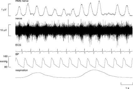
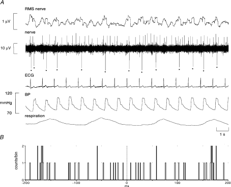
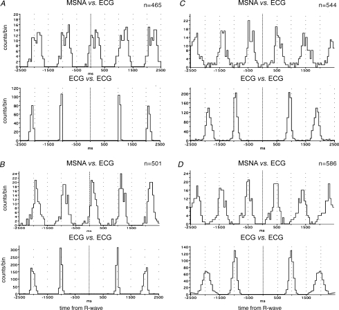
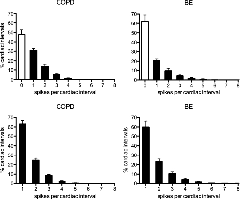
Elevated muscle sympathetic nerve activity (MSNA) features in many cardiovascular diseases, but how this sympathoexcitation is brought about differs across pathologies. Unitary recordings from post-ganglionic muscle vasoconstrictor neurones in human subjects have shown that the augmented MSNA in the obstructive sleep apnoea syndrome (OSAS) is associated with an increase in firing probability and mean firing rate, and an increase in multiple within-burst firing. Here we characterize the firing properties of muscle vasoconstrictor neurones in patients with chronic obstructive pulmonary disease (COPD), who are chronically asphyxic. We tested the hypothesis that this elevated chemical drive would shift the firing pattern from that seen in healthy subjects to that seen in OSAS. The mean firing probability (52%) and mean firing rate (0.92 Hz) of 17 muscle vasoconstrictor neurones recorded in COPD were comparable to those previously recorded in OSAS (51% and 0.96 Hz), but significantly higher than those recorded in a group of healthy subjects with high levels of resting MSNA (35% and 0.33 Hz). In COPD single neurones fired once in 63% of cardiac intervals, comparable to OSAS (59%), but significantly lower than in the healthy group (78%). Conversely, single neurones fired twice in 25% of cardiac intervals, similar to OSAS (27%), but significantly higher than in the healthy group (18%). We conclude that the chronic asphyxia associated with COPD results in an increase in the firing probability and mean firing frequency of muscle vasoconstrictor neurones and causes a shift towards multiple firing, reflecting an increase in central muscle vasoconstrictor drive.
Figures
References
-
- Carlsson JT, Hedner J, Elam M, Ejnell H, Sellgren J, Wallin BG. Augmented resting sympathetic activity in awake patients with obstructive sleep apnea. Chest. 1993;103:1763–1768. - PubMed
-
- Carlsson JT, Hedner J, Sellgren J, Elam M, Wallin BG. Depressed baroreflex sensitivity in patients with obstructive sleep apnea. Am J Respir Crit Care Med. 1996;154:1490–1496. - PubMed
-
- Cremona G, Higenbottam TW, Bower EA, Wood AM, Stewart S. Hemodynamic effects of basal and stimulated release of endogenous nitric oxide in isolated human lungs. Circulation. 1999;100:1316–1321. - PubMed
-
- De Troyer A, Leeper JB, McKenzie DK, Gandevia SC. Neural drive to the diaphragm in patients with severe COPD. Am J Respir Crit Care Med. 1997;155:1335–1340. - PubMed
-
- Elam M, Macefield VG. Multiple firing of single muscle vasoconstrictor neurons during cardiac dysrhythmias in human heart failure. J Appl Physiol. 2001;91:717–724. - PubMed
Publication types
MeSH terms
LinkOut - more resources
Full Text Sources
Medical