. 2010 Jan 5;3(103):ra1.
doi: 10.1126/scisignal.2000551.

Impaired alpha(IIb)beta(3) integrin activation and shear-dependent thrombus formation in mice lacking phospholipase D1

Affiliations

Impaired alpha(IIb)beta(3) integrin activation and shear-dependent thrombus formation in mice lacking phospholipase D1

Margitta Elvers et al. Sci Signal. .

Abstract

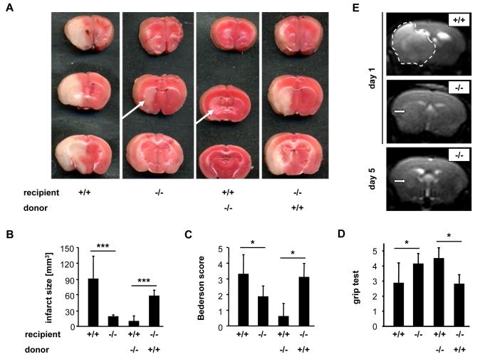
Platelet aggregation is essential for hemostasis but can also cause myocardial infarction and stroke. A key but poorly understood step in platelet activation is the shift of the principal adhesive receptor, alpha(IIb)beta(3) integrin, from a low- to high-affinity state for its ligands, a process that enables adhesion and aggregation. In response to stimulation of heterotrimeric guanosine triphosphate-binding protein or immunoreceptor tyrosine-based activation motif-coupled receptors, phospholipases cleave membrane phospholipids to generate lipid and soluble second messengers. An essential role in platelet activation has been established for phospholipase C (PLC) but not for PLD and its product phosphatidic acid. Here, we report that platelets from Pld1(-/-) mice displayed impaired alpha(IIb)beta(3) integrin activation in response to major agonists and defective glycoprotein Ib-dependent aggregate formation under high shear conditions. These defects resulted in protection from thrombosis and ischemic brain infarction without affecting tail bleeding times. These results indicate that PLD1 may be a critical regulator of platelet activity in the setting of ischemic cardiovascular and cerebrovascular events.


Figures

Fig. 1
Fig. 1
Impaired integrin αIIbβ3 activation in Pld1−/− platelets. (A) Washed blood from wild-type and Pld1−/− mice was incubated for 15 min with the indicated agonists in the presence of FITC-conjugated anti-mouse P-selectin antibody. The cells were gated by FSC and SSC characteristics. Data shown are mean fluorescence intensities (MFI) ± SD (n = 5 sets of cells per group). (B) Washed platelets were incubated with 50 μg/ml human Alexa-488 labeled fibrinogen and stimulated with PAR-4 peptide (NH2-AYPGKF) or CRP. Data shown are MFI ± SD of one representative experiment (n ≥ 4 sets of platelets per group. (C) Washed platelets were activated with 0.02 u/ml thrombin or 20 μg/ml for 2 min at 37°C under stirring conditions and fixed for 2 hours. Concentrations of ATP in the supernatant were determined using a bioluminescence assay kit. (D) Washed platelets were incubated with vehicle or 1 mM L-α-phosphatidic acid and analyzed as described in (B). (E) To assess aggregation, platelets from wild-type (black line) and Pld1−/− (gray line) mice were activated with the indicated agonists. Light transmission was recorded on a Fibrintimer 4 channel aggregometer over 10 min and was calculated as arbitrary units with 100% transmission adjusted with plasma. The results shown are representative of 3 individual experiments. 1 U/ml thrombin represents 11.4 nmol/L active protein.
Fig. 2
Fig. 2
Defective aggregate formation of Pld1−/− platelets on collagen at high shear. Whole blood was perfused over a collagen-coated (0.2 mg/ml coating) surface with the indicated shear rates, and then washed with Tyrode’s buffer for the same perfusion time. Perfusion times were 10 min (150 s−1) or 4 min (1000 s−1 and 1700 s−1). (A) Representative phase-contrast images at the end of the perfusion period. Scale bar, 100 μm. (B and C) Mean surface coverage (left) (B) and (C) relative platelet deposition as measured by integrated fluorescence intensity per visual field (right). All bar graphs depict mean values ± SD (n ≥ 3 mice each).
Fig. 3
Fig. 3
Pld1−/− platelets fail to firmly adhere to vWF under flow and display reduced coagulant activity. Whole blood was perfused over immobilized murine vWF with the indicated shear rates, and then washed with Tyrode’s buffer for the same perfusion time. (A and B) Adhesive platelets on the vWF-coated surface were counted after 100 s of blood perfusion (A) or at the end of the 4 min blood perfusion and washing step, reflecting firmly adhered platelets (B). All bar graphs depict mean values ± SD (n ≥ 3 mice each). (C and D) Whole blood was perfused over a collagen-coated (0.2 mg/ml) surface at a shear rate of 1000 s−1 or 1700 s−1 for 4 min. Adherent platelets were stained with 0.25 μg/ml OG-annexin A5. Representative phase-contrast (top) and fluorescence images (bottom) of the experiments performed at a shear rate of 1000 s−1 (C). Scale bar 100 μm. Mean relative amount of annexin A5 positive platelets ± SD (n ≥ 3 mice) for the indicated shear rates (D).
Fig. 4
Fig. 4
Reduced thrombus stability of Pld1−/− platelets in vivo. (A) Lethal pulmonary embolization after injection of collagen and epinephrine in anesthetized wild-type (+/+) and Pld1−/− (−/−) mice. Survivor rate upon injection of 700 μg collagen and 60 μg epinephrine per kg bodyweight (left). Representative pictures of lung sections (right). Note the large number of obstructed vessels in the wild-type lung section. (B) The right carotid artery of the indicated mice was injured by topical application of 15% ferric chloride and time to irreversible occlusion was determined with a Doppler flowmeter. Each symbol represents one individual mouse. (C to F) Mechanical injury of the abdominal aorta of wild-type (+/+) and Pld1−/− (−/−) mice was performed and blood flow was monitored with a Doppler flowmeter. Representative time course of blood flow in wild-type (black line) or Pld1−/− (gray line) aortas (C). Time to final occlusion, each symbol represents one individual mouse (D). Overall outcome for aortic injury. % distribution of irreversible occlusion (black), instable occlusion (light gray) and no occlusion (dark gray) (E). Representative cross-sections of the aorta 30 min after injury (F). Bars represent 200 μm. (G) Tail bleeding times for wild-type and Pld1−/− mice. Each symbol represents one animal.
Fig. 5
Fig. 5
Pld1−/− mice are protected from cerebral ischemia. (A) Representative images of three corresponding coronal sections from wild-type (+/+), Pld1−/− (−/−), wild-type mice transplanted with Pld1−/− bone marrow, and Pld1−/− mice transplanted with wild-type bone marrow stained with TTC 24 hours after tMCAO. (B) Brain infarct volumes. (C and D) Neurological Bederson score and grip test assessed at day 1 following tMACO of the mice mentioned in A. (E) Coronal T2-w MR brain imaging shows a large hyperintense ischemic lesion at day 1 after tMCAO in a wild-type mouse (top panel, demarcated by dotted line), but only a small infarct in a Pld1−/− mouse (middle panel, white arrow), and the T2 hyperintensity decreases by day 5 subsequent to infarct maturation (lower panel, white arrow). Hypointense areas indicating intracerebral hemorrhage were not seen in Pld1−/− mice, demonstrating that PLD1 deficiency does not increase the risk of hemorrhagic transformation, even at advanced stages of infarct development. Data are mean ± SD (n = 10 mice per group).

Similar articles

Cited by

References

    1. Ruggeri ZM. Platelets in atherothrombosis. Nat. Med. 2002;8:1227–1234. - PubMed
    1. Bhatt DL, Topol EJ. Scientific and therapeutic advances in antiplatelet therapy. Nat. Rev. Drug Discov. 2003;2:15–28. - PubMed
    1. Stoll G, Kleinschnitz C, Nieswandt B. Molecular mechanisms of thrombus formation in ischemic stroke: novel insights and targets for treatment. Blood. 2008;112:3555–3562. - PubMed
    1. Savage B, mus-Jacobs F, Ruggeri ZM. Specific synergy of multiple substrate-receptor interactions in platelet thrombus formation under flow. Cell. 1998;94:657–666. - PubMed
    1. Ozaki Y, Asazuma N, Suzuki-Inoue K, Berndt C. Platelet GPIb-IX-V-dependent signaling. J. Thromb. Haemost. 2005;3:1745–1751. - PubMed

MeSH terms