

Radiological and pathological size estimations of pure ductal carcinoma in situ of the breast, specimen handling and the influence on the success of breast conservation surgery: a review of 2564 cases from the Sloane Project
- PMID: 20051953
- PMCID: PMC2816666
- DOI: 10.1038/sj.bjc.6605513
Radiological and pathological size estimations of pure ductal carcinoma in situ of the breast, specimen handling and the influence on the success of breast conservation surgery: a review of 2564 cases from the Sloane Project
Abstract
Background: The Sloane Project, an audit of UK screen-detected non-invasive carcinomas and atypical hyperplasias of the breast, has accrued over 5000 cases in 5 years; with paired radiological and pathological data for 2564 ductal carcinoma in situ (DCIS) cases at the point of this analysis. We have compared the radiological estimate of DCIS size with the pathological estimate of DCIS size. We have correlated these sizes with histological grade, specimen-handling methods, particularly the use of specimen slice radiographs, and the success or failure of breast-conserving surgery (BCS).
Methods: The Sloane Project database was interrogated to extract information on all patients diagnosed with DCIS with complete radiological and pathological data on the size of DCIS, nuclear grade, specimen handling (with particular reference to specimen radiographs) and whether primary BCS was successful or whether the patient required further conservation surgery or a mastectomy.
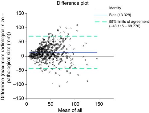
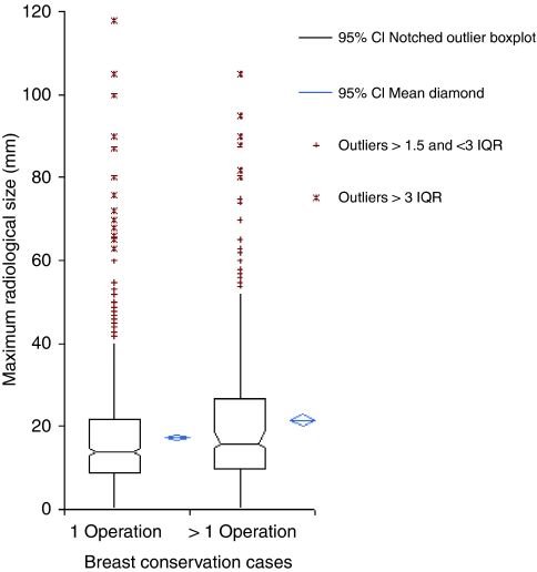
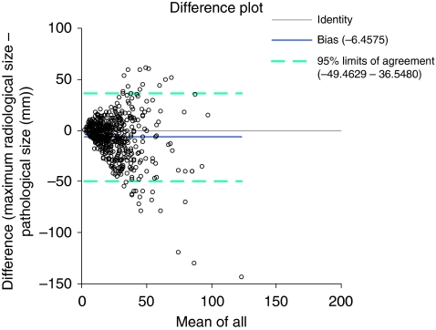
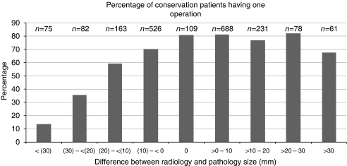
Results: Of 2564 patients in the study, 2013 (79%) had attempted BCS and 1430 (71%) had a successful single operation. Of the 583 BCS patients who required further surgery, 65% had successful conservation and 97% of them after a single further operation. In successful one-operation BCS patients, there was a close agreement between radiological and pathological DCIS size with radiology tending to marginally overestimate the disease extent. In multiple-operation BCS, radiology underestimated DCIS size in 59% of cases. The agreement between pathological and radiological size of DCIS was poor in mastectomies but was improved by specimen slice radiography, suggesting specimen-handling techniques as a cause.
Conclusion: In 30% of patients undergoing BCS for DCIS, preoperative imaging underestimates the extent of disease resulting in a requirement for further surgery. This has implications for the further improvement of preoperative imaging and non-operative diagnosis of DCIS so that second operations are reduced to a minimum.
Figures
References
-
- Altman DG, Bland JM (1983) Measurement in medicine: the Analysis of Method Comparison Studies. Statistician 32: 307–317
-
- Asjoe FT, Altinas S, Colpaert C, Marck EV, Vermorken JB, Tjalma WA (2007) The value of the Van Nuys prognostic index in ductal carcinoma in situ of the breast: a retrospective analysis. Breast J 13: 359–367 - PubMed
-
- Bagnall MJ, Evans AJ, Wilson AR, Pinder SE, Denley H, Geraghty JG, Ellis IO (2001) Predicting invasion in mammographically detected microcalcification. Clin Radiol 56: 828–832 - PubMed
-
- Bland JM, Altman DG (1986) Statistical methods for assessing agreement between two methods of clinical measurement. Lancet 1(8476): 307–310 - PubMed
-
- Chakrabarti J, Evans AJ, James J, Ellis IO, Pinder SE, Macmillan RD (2006) Accuracy of mammography in predicting histological extent of ductal carcinoma in situ (DCIS). EJSO 32: 1089–1092 - PubMed
Publication types
MeSH terms
LinkOut - more resources
Full Text Sources
Medical
