. 2010 Jan 19;102(2):436-46.
doi: 10.1038/sj.bjc.6605516. Epub 2010 Jan 5.

EWS-Oct-4B, an alternative EWS-Oct-4 fusion gene, is a potent oncogene linked to human epithelial tumours

Affiliations

EWS-Oct-4B, an alternative EWS-Oct-4 fusion gene, is a potent oncogene linked to human epithelial tumours

S Kim et al. Br J Cancer. .

Abstract

Background: Characterisation of EWS-Oct-4 translocation fusion product in bone and soft-tissue tumours revealed a chimeric gene resulting from an in-frame fusion between EWS (Ewing's sarcoma gene) exons 1-6 and Oct-4 exons 1-4. Recently, an alternative form of the fusion protein between the EWS and Oct-4 genes, named EWS-Oct-4B, was reported in two types of epithelial tumours, a hidradenoma of the skin and a mucoepidermoid carcinoma of the salivary glands. As the N-terminal and POU domains of the EWS-Oct-4 and EWS-Oct-4B proteins are not structurally identical, we decided to investigate the functional consequences of the EWS-Oct-4B fusion.

Methods: In this report, we have characterised the EWS-Oct-4B fusion protein. To investigate how the EWS-Oct-4B protein contributes to tumourigenesis in human cancers, we analysed its DNA-binding activity, subcellular localisation, transcriptional activation behaviour, and oncogenic properties.

Results: We found that this new chimeric gene encodes a nuclear protein that binds DNA with the same sequence specificity as the parental Oct-4 protein or the fusion EWS-Oct-4 protein. We show that the nuclear localisation signal of EWS-Oct-4B is dependent on the POU DNA-binding domain, and we identified a cluster of basic amino acids, (269)RKRKR(273), in the POU domain that specifically mediates the nuclear localisation of EWS-Oct-4B. Comparison of the properties of EWS-Oct-4B and EWS-Oct-4 indicated that EWS-Oct-4B is a less-potent transcriptional activator of a reporter construct carrying the Oct-4-binding sites. Deletion analysis of the functional domains of EWS-Oct-4B revealed that the EWS N-terminal domain (NTD)(B), POU, and C-terminal domain (CTD) are necessary for its full transactivation potential. Despite its reduced activity as a transcriptional activator, EWS-Oct-4B regulated the expression of fgf-4 (fibroblast growth factor-4) and nanog, which are potent mitogens, as well as of Oct-4 downstream target genes, the promoters of which contain potential Oct-4-binding sites. Finally, ectopic expression of EWS-Oct-4B in Oct-4-null ZHBTc4 ES cells resulted in increased tumourigenic growth potential in nude mice.

Conclusion: These results suggest that the oncogenic effect of the t(6;22) translocation is due to the EWS-Oct-4B chimeric protein, and that alternative fusion of the EWS amino terminal domain to the Oct-4 DNA-binding domain produces another transforming chimeric product in human epithelial tumours.


Figures

Figure 1
Figure 1
Structural comparison of EWS-Oct-4 alternative forms. Two different forms of the EWS-Oct-4 protein (EWS-Oct-4 and EWS-Oct-4B) and the human Oct-4 isoforms (Oct-4A and Oct-4B) are represented schematically. Amino acid (aa) positions are indicated above and below the schematic representing the proteins. In the case of EWS-Oct-4, the first 193 aa (residues 1–193) of EWS are fused to the truncated coding sequence (residues 123–360) of Oct-4A through an additional six aa sequence. This six aa sequence is encoded by the normal intron 6 of EWS in the fusion transcript that lacks the first 122 aa (residues 1–122) present in Oct-4A. In contrast, in the case of EWS-Oct-4B, the first 174 aa (residues 1–174) of EWS were fused to the POU DNA-binding domain (residues 136–360) of Oct-4A, in which neither the additional six aa sequence nor the N-terminal domain of Oct-4A is retained in EWS-Oct-4B. Functionally important domains of the Oct-4 isoforms and EWS-Oct-4 chimeras are indicated: NTDA, the N-terminal domain of human Oct-4A; POUA, POU DNA-binding domain (total 156 amino acids) of human Oct-4A; CTD, C-terminal domain of Oct-4; NTDB, N-terminal domain of human Oct-4B; POUB, POU DNA-binding domain (total 154 amino acids) of human Oct-4B; EWS (NTD), EWS N-terminal domain (residues 1–193) of EWS-Oct-4; Extra 6 aa, extra six amino acids; N, truncated N-terminal domain of Oct-4; EWS (NTD)B, EWS N-terminal domain (residues 1–174) of EWS-Oct-4B.
Figure 2
Figure 2
Mapping of the nuclear localisation signal of EWS-Oct-4B to the POUB DNA-binding domain. (A) Subcellular localisation of EWS-Oct-4B. Cells (293T) grown on coverslips were transfected with mammalian expression vectors encoding (a) EGFP or (b) EWS-Oct-4B-EGFP. The subcellular localisation of the EWS-Oct-4B protein was determined by monitoring the location of the green fluorescence. (B) Schematic diagram showing EWS-Oct-4B truncation mutants of GST-EGFP fusion proteins. Subcellular localisation of the indicated truncation mutants was determined by monitoring the location of green fluorescence, and is indicated as N (nuclear localisation) or C (cytoplasmic localisation). (C) Subcellular distribution of EWS-Oct-4B deletion mutants. 293T cells were grown on coverslips under low-density conditions and transfected with expression plasmids for the indicated GST-EGFP-EWS-Oct-4B deletion mutants. The cells were fixed with an acetone/methanol mixture and EGFP was analysed by fluorescence microscopy.
Figure 3
Figure 3
Comparison of transactivation potential by EWS-Oct-4B. Cells (293T) were cotransfected with expression vectors encoding the indicated amounts of EWS-Oct-4 (grey bars) or EWS-Oct-4B (black bars), the pOct-4(10 × ) TATA luc reporter plasmid, and the Renilla luciferase (top panel). Reporter activity was normalised with Renilla luciferase activity to correct for different transfection efficiencies. Fold induction is expressed relative to the empty expression vector. Each transfection was performed at least thrice independently and the mean values are plotted with their standard errors (±s.e., vertical bars). Extracts for luciferase assays were resolved using 12% SDS–PAGE, transferred to a PVDF membrane, and immunoblotted with anti-Oct-4 (C-20) (middle panel) or anti-EGFP (bottom panel) antibodies, as indicated.
Figure 4
Figure 4
DNA-binding property of the EWS-Oct-4B chimera. (A) Immunoblot analysis of EWS-Oct-4B and EWS-Oct-4 to quantify GST fusion proteins. The GST-EWS-Oct-4 fusion proteins used in EMSAs were fractionated on 10% SDS–PAGE and visualised by western blotting with an anti-GST antibody (B-14, Santa Cruz Biotechnology). (B) EMSAs of the DNA-binding properties of EWS-Oct-4B and EWS-Oct-4. The EMSAs were performed using recombinant GST (lane 1, 0.15 μg; lane 2, 0.45 μg; lane 3, 1.35 μg), GST-EWS-Oct-4 (lane 4, 0.15 μg; lane 5, 0.45 μg; lane 6; 1.35 μg), or GST-EWS-Oct-4B (lane 7, 0.15 μg; lane 8, 0.45 μg; lane 9, 1.35 μg), and radiolabeled probe, as described in Materials and Methods. The recombinant proteins used in each EMSA are indicated above the gel. Protein–DNA complexes were resolved on non-denaturing 4% polyacrylamide (acrylamide:bisacrylamide ratio, 37 : 1) gels run at 4°C in 0.5 × TBE (44.5 mM Tris-HCl, 44.5 mM boric acid, 1 mM EDTA). The positions of free probe and protein–DNA complexes are indicated.
Figure 5
Figure 5
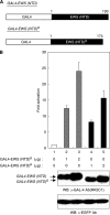
Transactivation potential of EWS (NTD)B. (A) Schematic representation of the GAL4-fusion expression plasmids used in this study. The expression vectors driving the production of GAL4-EWS (NTD) or GAL4-EWS (NTD)B are shown. GAL4, GAL4 DNA-binding domain; EWS (NTD), EWS N-terminal domain of EWS-Oct-4; EWS (NTD)B, EWS N-terminal domain of EWS-Oct-4B. (B) Comparison of transactivation potential of EWS (NTD)B. The reporter plasmid, 5 × Gal4 TATA luc, was cotransfected with GAL4-EWS (NTD) or GAL4-EWS (NTD)B into 293T cells (top panel). Luciferase activity was expressed as fold activation relative to the basal level observed with the reporter plasmid and the GAL4 DNA-binding domain alone (lane 1). Each transfection was performed independently at least thrice and the mean values are plotted with their standard errors (±s.e., vertical bars). Extracts for luciferase assays were resolved using 12% SDS–PAGE, transferred to a PVDF membrane, and immunoblotted with anti-GAL-4 (RK5C1) (middle panel) or anti-EGFP (bottom panel) antibodies as indicated.
Figure 6
Figure 6
Functional regions of EWS-Oct-4B. (A) Transcriptional properties of EWS-Oct-4B deletion mutants. Shown on the left are schematic representations of EWS-Oct-4B deletion constructs. A reporter plasmid, pOct-4(10 × ) TATA luc, was cotransfected into 293T cells with various flag-tagged EWS-Oct-4B mutants. Relative transcriptional activation values are shown on the right as mean increases±s.e. relative to a value of 100% for transfection of EWS-Oct-4B. The results are the means of three independent experiments performed in duplicate. (B) Immunoblot analysis showing expression of the EWS-Oct-4B deletion mutants in transiently transfected cells. Total cell lysates were fractionated by 12% SDS–PAGE and visualised by western blotting with anti-Flag (M2, Sigma, top panel) or anti-EGFP (Invitrogen Molecular Probes, bottom panel) antibodies. The positions of pre-stained molecular weight markers (New England Biolabs, Hitchin, UK) are indicated to the left (kDa). Lane 1, empty vector; lane 2, EWS-Oct-4B; lane 3, EWS-Oct-4B (ΔEWS); lane 4, EWS-Oct-4 (ΔCTD); and lane 5, EWS-Oct-4B (V313P).
Figure 7
Figure 7
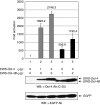
Transcriptional activation of Oct-4 downstream target genes by EWS-Oct-4B in vivo. (A) Schematic representation of the expression vectors. Expression vector pCAG-IP/EWS-Oct-4B-EGFP corresponds to EWS-Oct-4B fused to EGFP. The pCAG-IP/EGFP expression vector was used as a control. The CAG expression units (CAG) are indicated by shaded boxes, EWS-Oct-4B is represented by an open box, and EGFP is indicated by solid boxes. (B) Immunoblot analysis of EWS-Oct-4B expression in stably transfected ZHBTc4 ES cells. Total cell lysates (60 μg protein) were fractionated by 12% SDS–PAGE and visualised by western blotting with anti-EGFP (Invitrogen Molecular Probes, top panel) or anti-GAPDH (V-18, Santa Cruz Biotechnology, lower panel) antibodies. (C) Induction of Oct-4 downstream target genes by EWS-Oct-4B in vivo. Northern blot analyses of fgf-4 and nanog mRNAs were performed in ZHBTc4 ES cells expressing vector (lane 1) or EWS-Oct-4B fusion proteins (lane 2). Total RNA was fractionated on a 6% formaldehyde–1.5% agarose gel, transferred to a nylon membrane, and probed using mouse fgf-4 (upper panel) or nanog (second panel) cDNAs, as described in Materials and Methods. Ethidium bromide (EtBr) staining of the agarose gel used for northern blotting shows that equal amounts of total RNA were loaded in each lane (lower panel). The stable cell lines from which the total RNAs used in the northern blot analysis were derived are shown above the panel.
Figure 8
Figure 8
Characterisation of the effects of ZHBTc ES cells expressing EWS-Oct-4B in nude mice. (A) Effects of EWS-Oct-4B expression on the development of tumours in nude mice. Approximately 0.6 × 10⁷ Oct-4-null ZHBTc4 ES cells expressing EWS-Oct-4B-EGFP or EGFP, were suspended in 100 μl PBS and injected into 5-week-old Balb/c athymic nude mice. Tumour development was observed and photographs were taken 26 days after injection. (B) Weight of tumours from nude mice injected with Oct-4-null ZHBTc ES cells expressing EWS-Oct-4B. Tumour weight in individual animals was measured at 26 days after injection (n=6) and is plotted as mean increases±s.e.

Similar articles

Cited by

References

    1. Aasen T, Raya A, Barrero MJ, Garreta E, Consiglio A, Gonzalez F, Vassena R, Bilic J, Pekarik V, Tiscornia G, Edel M, Boue S, Belmonte JC (2008) Efficient and rapid generation of induced pluripotent stem cells from human keratinocytes. Nat Biotechnol 26: 1276–1284 - PubMed
    1. Ambrosetti DC, Basilico C, Dailey L (1997) Synergistic activation of the fibroblast growth factor 4 enhancer by Sox2 and Oct-3 depends on protein-protein interactions facilitated by a specific spatial arrangement of factor binding sites. Mol Cell Biol 17: 6321–6329 - PMC - PubMed
    1. Botquin V, Hess H, Fuhrmann G, Anastassiadis C, Gross MK, Vriend G, Scholer HR (1998) New POU dimer configuration mediates antagonistic control of an osteopontin preimplantation enhancer by Oct-4 and Sox-2. Genes Dev 12: 2073–2090 - PMC - PubMed
    1. Breeuwer M, Goldfarb DS (1990) Facilitated nuclear transport of histone H1 and other small nucleophilic proteins. Cell 60: 999–1008 - PubMed
    1. Brehm A, Ohbo K, Scholer H (1997) The carboxy-terminal transactivation domain of Oct-4 acquires cell specificity through the POU domain. Mol Cell Biol 17: 154–162 - PMC - PubMed

Publication types

MeSH terms

Substances