

Frequent genetic differences between matched primary and metastatic breast cancer provide an approach to identification of biomarkers for disease progression
- PMID: 20051991
- PMCID: PMC2987320
- DOI: 10.1038/ejhg.2009.230
Frequent genetic differences between matched primary and metastatic breast cancer provide an approach to identification of biomarkers for disease progression
Abstract
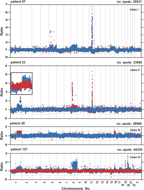
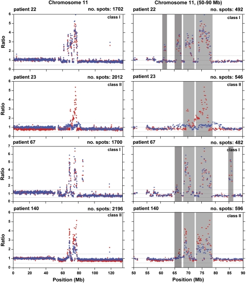
Breast cancer is a major cause of morbidity and mortality in women and its metastatic spread is the principal reason behind the fatal outcome. Metastasis-related research of breast cancer is however underdeveloped when compared with the abundant literature on primary tumors. We applied an unexplored approach comparing at high resolution the genomic profiles of primary tumors and synchronous axillary lymph node metastases from 13 patients with breast cancer. Overall, primary tumors displayed 20% higher number of aberrations than metastases. In all but two patients, we detected in total 157 statistically significant differences between primary lesions and matched metastases. We further observed differences that can be linked to metastatic disease and there was also an overlapping pattern of changes between different patients. Many of the differences described here have been previously linked to poor patient survival, suggesting that this is a viable approach toward finding biomarkers for disease progression and definition of new targets useful for development of anticancer drugs. Frequent genetic differences between primary tumors and metastases in breast cancer also question, at least to some extent, the role of primary tumors as a surrogate subject of study for the systemic disease.
Figures
References
-
- Eccles S, Paon L, Sleeman J. Lymphatic metastasis in breast cancer: importance and new insights into cellular and molecular mechanisms. Clin Exp Metastasis. 2007;24:619–636. - PubMed
-
- Bild AH, Potti A, Nevins JR. Linking oncogenic pathways with therapeutic opportunities. Nat Rev. 2006;6:735–741. - PubMed
-
- Driouch K, Landemaine T, Sin S, Wang S, Lidereau R. Gene arrays for diagnosis, prognosis and treatment of breast cancer metastasis. Clin Exp Metastasis. 2007;24:575–585. - PubMed
-
- Sotiriou C, Piccart MJ. Taking gene-expression profiling to the clinic: when will molecular signatures become relevant to patient care. Nat Rev. 2007;7:545–553. - PubMed
Publication types
MeSH terms
Substances
Grants and funding
LinkOut - more resources
Full Text Sources
Medical
