. 2010 Jan 5;5(1):e8563.
doi: 10.1371/journal.pone.0008563.

Presynaptic external calcium signaling involves the calcium-sensing receptor in neocortical nerve terminals

Affiliations

Presynaptic external calcium signaling involves the calcium-sensing receptor in neocortical nerve terminals

Wenyan Chen et al. PLoS One. .

Abstract

Background: Nerve terminal invasion by an axonal spike activates voltage-gated channels, triggering calcium entry, vesicle fusion, and release of neurotransmitter. Ion channels activated at the terminal shape the presynaptic spike and so regulate the magnitude and duration of calcium entry. Consequently characterization of the functional properties of ion channels at nerve terminals is crucial to understand the regulation of transmitter release. Direct recordings from small neocortical nerve terminals have revealed that external [Ca(2+)] ([Ca(2+)](o)) indirectly regulates a non-selective cation channel (NSCC) in neocortical nerve terminals via an unknown [Ca(2+)](o) sensor. Here, we identify the first component in a presynaptic calcium signaling pathway.

Methodology/principal findings: By combining genetic and pharmacological approaches with direct patch-clamp recordings from small acutely isolated neocortical nerve terminals we identify the extracellular calcium sensor. Our results show that the calcium-sensing receptor (CaSR), a previously identified G-protein coupled receptor that is the mainstay in serum calcium homeostasis, is the extracellular calcium sensor in these acutely dissociated nerve terminals. The NSCC currents from reduced function mutant CaSR mice were less sensitive to changes in [Ca(2+)](o) than wild-type. Calindol, an allosteric CaSR agonist, reduced NSCC currents in direct terminal recordings in a dose-dependent and reversible manner. In contrast, glutamate and GABA did not affect the NSCC currents.

Conclusions/significance: Our experiments identify CaSR as the first component in the [Ca(2+)](o) sensor-NSCC signaling pathway in neocortical terminals. Decreases in [Ca(2+)](o) will depress synaptic transmission because of the exquisite sensitivity of transmitter release to [Ca(2+)](o) following its entry via voltage-activated Ca(2+) channels. CaSR may detects such falls in [Ca(2+)](o) and increase action potential duration by increasing NSCC activity, thereby attenuating the impact of decreases in [Ca(2+)](o) on release probability. CaSR is positioned to detect the dynamic changes of [Ca(2+)](o) and provide presynaptic feedback that will alter brain excitability.


Conflict of interest statement

Competing Interests: R.H.D., P.D., and M.R. have filed a use-patent for Calindol. This does not alter adherence to all the PLoS ONE policies on sharing data and materials, as detailed online in the guide for authors.

Figures

Figure 1
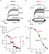
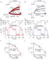
Figure 1. Loss of function CaSR mutation reduces NSCC current sensitivity to [Ca2+]o.
Cell- attached recordings were made from (A) CaSR+/+ and (B) CaSR+/− terminals and the Ca2+ in the bath solution applied to terminals varied between 6 µM and 60 mM as indicated. Step depolarizations (−40 to 110 mV relative to resting membrane potential) were made every 5 seconds. Average current traces (n = 8−15) are shown for each bath [Ca2+] at steady-state for two exemplar recordings. Note that outward currents elicited with 0.6 and 6 mM Ca2+ were proportionately larger in the heterozygote than in the wild-type recording. C) timecourse of NSCC current amplitude (measured at the end of the depolarizing step) in the same CaSR+/+ (filled circles, left axis) and CaSR+/− (open circles, right axis) terminals as bath [Ca2+] was increased (upper trace). Steady state amplitude was reached in 5–10 s for both CaSR+/+ and CaSR+/− genotypes. Axes were scaled to span the current amplitudes measured between with bath [Ca2+] between 6 µM and 60 mM. D, the concentration-effect relationship for both CaSR genotypes shows that wild-type terminals exhibited higher affinity for Ca2+ (p = 0.032). NSCC currents were normalized for each terminal by measuring the difference between the NSCC current and the 60 mM Ca2+-elicited NSCC current and dividing this by the difference between the NSCC currents elecited by 6 µM and 60 mM Ca2+. The curves represent mean±SEM of 7 and 6 recordings for CaSR+/+ and CaSR+/−, respectively. The curves were fit to the average data points resulting in Hill coefficients of 0.77 for both genotypes and IC50s of 1.6±0.2 mM and 1.1±0.07 mM for CaSR+/− and CaSR+/+, respectively.
Figure 2
Figure 2. CaSR is present in nerve terminals of neocortex.
A, immunoblot of synaptosomes and whole brain show 160 kDa bands with anti-CaSR antibody . Positive control (HEK CaR) shows 140 and 160 kDa bands (glycosylated and unglycosylated forms) in CaSR- transfected HEK cells and no signal in untransfected HEK cells (HEK Con). B, acutely isolated nerve terminals (synaptosomes) identified using the synaptophysin antibody (red). C, CaSR identified with polyclonal antibody “4641” (green). D, superimposition of B and C shows that CaSR and synaptophysin are co-localized.
Figure 3
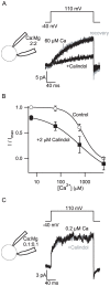
Figure 3. Calindol facilitates inhibition of NSCC currents by extracellular Ca2+.
A, exemplar traces show NSCC currents reversibly inhibited by addition of Calindol (10 µM) to bath solution. Traces were recorded in cell-attached mode following 200 ms step depolarization with 60 µM Ca2+ and 0 Mg2+ in the bath and 2 mM Ca2+ and 2 mM Mg2+ in the pipette solution (inset). The substantial inhibition of NSCC current was reversed following washout (gray trace). B, the Ca2+ concentration-effect relationship was left-shifted by the allosteric CaSR agonist Calindol (2 µM). These data represent 11 synaptosome recordings, each normalized to the current observed in 6 µM Ca2+. C, average traces show NSCC currents unaffected by the addition of 10 µM Calindol (gray trace) to bath solution containing reduced [Ca2+]o. Traces were recorded in cell-attached recording following 200 ms step depolarization with 0.2 µM Ca2+ and 0 Mg2+ in the bath and 0.1 mM Ca2+ and 0.1 Mg2+ in the pipette solution (inset). Recordings were less stable at low divalent concentrations; traces are thus averages of 8 currents elicited with a 5 second duty cycle.
Figure 4
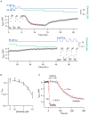
Figure 4. Kinetics and dose-dependence of Calindol inhibtion of NSCC currents in synaptosomes.
A, contiguous plot of NSCC current amplitude versus time showing timecourse of on and off kinetics for Calindol action in a cell-attached synaptosome recording. The outward current elicited by voltage step (200 ms, −40 to 110 mV) every 5 seconds is plotted against time. NSCC current was reversibly and reliably activated by reductions in bath [Ca2+] (0.06–6 mM; blue trace) and inhibited by Calindol (0.1–10 µM; green trace). The Ca2+ and Calindol axes are logarithmic and the absence of the green trace indicates Calindol concentration is zero. Calindol (10 µM) inhibited NSCC current after a 1–2 minute delay and thereafter blocked with mono-exponential time course. The current increased with an exponential time course following reduction of Calindol. B, Calindol inhibited NSCC currents in bath [Ca2+] of 60 µM with an IC50 6.3±1.1 µM on average (n = 4). Each recording was normalized by dividing the NSCC current amplitude by the NSCC current amplitude elicited by 60 µM bath [Ca2+] before Calindol was applied. Calindol was generally applied at higher concentrations first and the amplitude of the NSCC current increased after washout (closed triangle) compared to initial baseline (open circle). This presumably reflects a run-up phenomenon due to the long duration of these experiments. C, Calindol inhibition of NSCC current is biphasic (i.e. latency and monoexponential) whereas Ca2+ inhibition is well described by a single exponential. Two sections of the timecourse data in A displaying applications of 10 µM Calindol and 6 mM Ca2+ (just prior to Calindol application) were redrawn on expanded time axis and overlaid so that time zero corresponded to solution change. Both datasets are well-fit by single exponentials with Calindol decaying significantly more slowly (tau of 73 s for Calindol vs 6.6 s for Ca2+) and at a substantial latency.
Figure 5
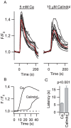
Figure 5. Calindol activation of CaSR expressed in HEK cells occurred with a greater latency than Ca2+ activation of CaSR.
A, application of Ca2+ (5 mM) and Calindol (10 µM) at time zero to CaSR-expressing HEK cells in 1 mM Ca2+ and 0 Mg2+ caused a transient increase in fluorescence (F) relative to basal level (F0) indicating an increase in [Ca2+]i. The black curves denote signal from 12 cells and the red curves indicate the average. There was a 5 minute delay between applications. B, average curves from A have been redrawn on the same time-expanded axis to compare effect latency of Calindol vs. Ca2+. C, histogram of latency of effect for Ca2+ and Calindol. The latency was significantly greater for activation by Calindol (23.7±1.2 s) than by Ca2+ alone (9.0±0.2 s) in the recordings from 22 cells (p<0.001).
Figure 6
Figure 6. Glutamate (10 µM) but not Calindol (5 µM) activated the mGluR1 expressed in HEK cells.
Application of glutamate (horizontal bars) caused a transient increase in fluorescence (F) relative to basal level (F0) indicating an increase in [Ca2+]i (left). The black curves denote signals from 35 cells while red curves indicate averages. Applications of test solutions were staggered by 10 min to allow for recovery. Calindol application (horizontal bar) to the same cells did not produce any change in F/F0 (middle), yet these cells remained responsive to glutamate (right).
Figure 7
Figure 7. The [Ca2+]o-modulated NSCC current in rat nerve terminals was unaffected by glutamate and GABA.
A, currents activated by step depolarizations (−40 to 110 mV relative to membrane potential) across a range of bath [Ca2+] (60 µM-6 mM, black traces) superimposed with those recorded during addition of 100 µM glutamate to the perfusate (red traces). Each trace represents an average of 10–20 currents at steady state solution conditions from the same cell-attached recording from a synaptosome. B, timecourse of NSCC current amplitude activated by the 200 ms voltage step every 5 seconds at three [Ca2+]o (upper trace). Same recording as A. Glutamate application is denoted by horizontal bar. C, normalized concentration-effect relationship for changes in [Ca2+]o from NSCC synaptosome recordings is unaffected by glutamate (n = 6). Currents represent NSCC current amplitude at end of voltage step relative to current before step divided by NSCC current amplitude with 60 µM Ca2+ in bath. D, currents activated by step depolarizations over a range of bath [Ca2+] (60 µM-6 mM, black traces) superimposed with those recorded during perfusion of 100 µM GABA (red traces). Each trace represents an average of 10–20 currents at steady state solution conditions from the same cell-attached recording from a synaptosome. E, timecourse of NSCC current amplitude from D at three [Ca2+]o (upper trace). GABA application is denoted by horizontal bar. E, normalized concentration-effect relationship for [Ca2+] o and NSCC amplitude in synaptosome recordings is unaffected by GABA (n = 3). Currents represent NSCC current amplitude at end of voltage step relative to current before step divided by NSCC current amplitude with 60 µM Ca2+ in bath.

Similar articles

Cited by

References

    1. Neher E, Sakaba T. Multiple roles of calcium ions in the regulation of neurotransmitter release. Neuron. 2008;59:861–872. - PubMed
    1. Bischofberger J, Geiger JR, Jonas P. Timing and efficacy of Ca2+ channel activation in hippocampal mossy fiber boutons. J Neurosci. 2002;22:10593–10602. - PMC - PubMed
    1. Li L, Bischofberger J, Jonas P. Differential Gating and Recruitment of P/Q-, N-, and R-Type Ca2+ Channels in Hippocampal Mossy Fiber Boutons. Journal of Neuroscience. 2007;27:13420–13429. - PMC - PubMed
    1. Geiger JR, Jonas P. Dynamic control of presynaptic Ca(2+) inflow by fast-inactivating K(+) channels in hippocampal mossy fiber boutons. Neuron. 2000;28:927–939. - PubMed
    1. Engel D, Jonas P. Presynaptic Action Potential Amplification by Voltage-Gated Na+ Channels in Hippocampal Mossy Fiber Boutons. Neuron. 2005;45:405–417. - PubMed

Publication types

MeSH terms