. 2010 Jan 5;5(1):e8560.
doi: 10.1371/journal.pone.0008560.

Myosin II motor proteins with different functions determine the fate of lamellipodia extension during cell spreading

Affiliations

Myosin II motor proteins with different functions determine the fate of lamellipodia extension during cell spreading

Venkaiah Betapudi. PLoS One. .

Abstract

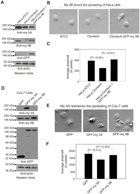
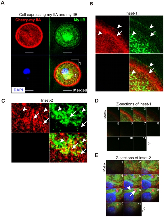
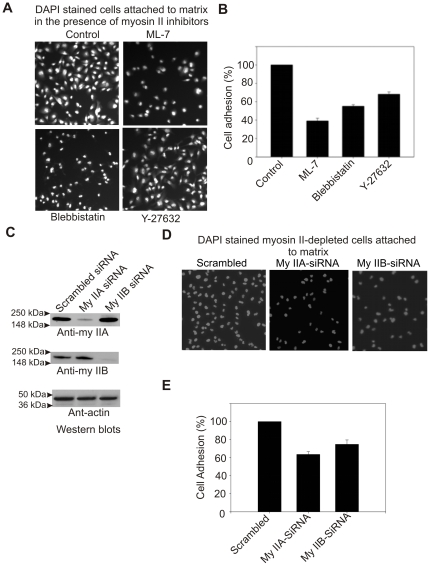
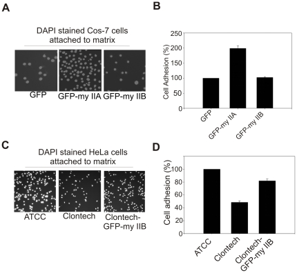
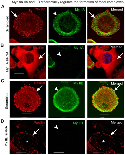
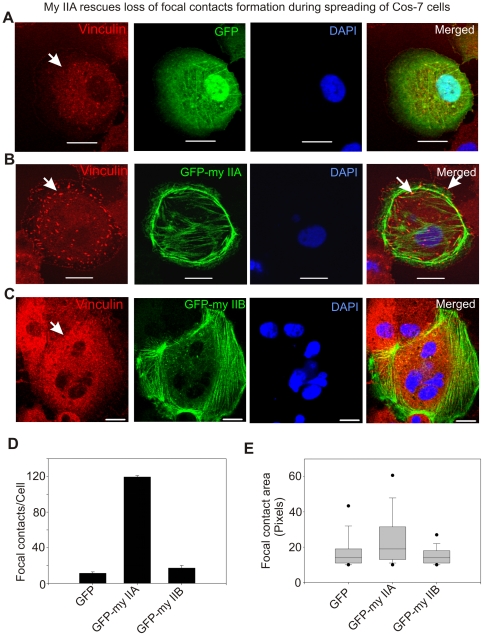
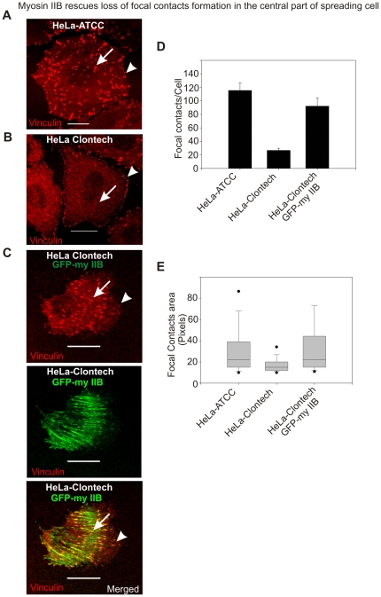
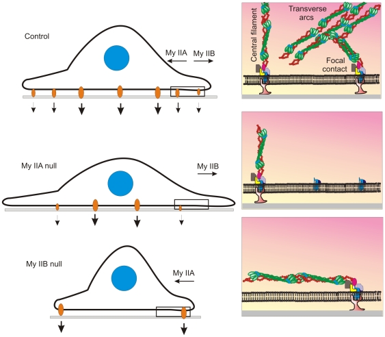
Non-muscle cells express multiple myosin-II motor proteins myosin IIA, myosin IIB and myosin IIC transcribed from different loci in the human genome. Due to a significant homology in their sequences, these ubiquitously expressed myosin II motor proteins are believed to have overlapping cellular functions, but the mechanistic details are not elucidated. The present study uncovered a mechanism that coordinates the distinctly localized myosin IIA and myosin IIB with unexpected opposite mechanical roles in maneuvering lamellipodia extension, a critical step in the initiation of cell invasion, spreading, and migration. Myosin IIB motor protein by localizing at the front drives lamellipodia extension during cell spreading. On the other hand, myosin IIA localizes next to myosin IIB and attenuates or retracts lamellipodia extension. Myosin IIA and IIB increase cell adhesion by regulating focal contacts formation in the spreading margins and central part of the spreading cell, respectively. Spreading cells expressing both myosin IIA and myosin IIB motor proteins display an organized actin network consisting of retrograde filaments, arcs and central filaments attached to focal contacts. This organized actin network especially arcs and focal contacts formation in the spreading margins were lost in myosin IIA cells. Surprisingly, myosin IIB cells displayed long parallel actin filaments connected to focal contacts in the spreading margins. Thus, with different roles in the regulation of the actin network and focal contacts formation, both myosin IIA and IIB determine the fate of lamellipodia extension during cell spreading.


Conflict of interest statement

Competing Interests: The author has declared that no competing interests exist.

Figures

Figure 1
Figure 1. Myosin IIA and IIB mediate cell membrane extension in opposite directions.
A) Western blots showing the expression of myosin II isoforms in HeLa cells. B) Myosin IIB favors lamellipodium extension during cell spreading. C) Graphical representation of the cell spread areas. Cells spread for 60 min were stopped by directly adding fixing buffer followed by staining with Alexa-Fluor conjugated wheat germ agglutinin to enhance cell margin contrast, and quantifying cell area using the ImageJ program (NIH) as described in Methods. For each presented bar, n = 350–450 cells quantified. The average spread area of HeLa-ATCC cell was considered as 100% to calculate the spread area of other cells. D) Western blots showing the expression of myosin II isoforms in COS-7 cells. E) Myosin IIA mediates lamellipodia retraction during cell spreading. F) Graphical representation of the cell spread areas. For each presented bar, n = 550–650 cells quantified. Bars represent standard errors, t-test. The average spread area of GFP alone expressing cell was considered as 100% to calculate the spread area of other cells.
Figure 2
Figure 2. Myosin II undergoes assembly regulation during cell spreading.
A) Myosin II assembly and construction of myosin II-ACD expression vectors. MDA-MB 231 cells were used for transient expression of myosin II-ACD. B) Spreading MDA-MB 231 cells transiently expressing myosin II-ACD. Images were collected after 60 min spread. C) Graphical representation of spreading areas of cells expressing myosin II-ACD. Spread areas of cells were measured as described under Fig. 1 legend. For each presented bar, n = 350–400 cells quantified. Bars represent standard errors, t-test. D) A schematic diagram showing the opposite roles of myosin IIA and IIB in extending lamellipodia during spreading.
Figure 3
Figure 3. Distinct localizations of myosin IIA and IIB in spreading MDA-MB 231 breast cancer cells.
A) Spreading cell transiently expressing cherry-myosin IIA was fixed and stained with myosin IIB antibody. A series of Z-sections were collected using confocal microscope. Inset-1 and inset-2 were drawn to study colocalizations of myosin IIA and IIB at the spreading margins and in the cytoplasm, respectively. B) and C) Blowup of a z-sections collected near the matrix. Arrow indicates cherry-myosin IIA and arrow-head shows endogenous myosin IIB. D) and E) Myosin IIA and IIB display distinct localization in the spreading margins and cytoplasm. A series of z-sections were collected using confocal microscope.
Figure 4
Figure 4. Myosin IIA and IIB involved in the regulation of cell attachment to matrix.
A) Pharmacological inhibition of myosin II impairs cell attachment to matrix. B) Graphical representation of cell adhesion impairment in the presence of myosin II inhibitors. DAPI stained nuclei on the images were quantified using ImageJ program (NIH) as a measure of cell number. The average number of cells attached in the control population was set as 100%. C) Western blots showing depletion of myosin II isoforms in siRNA electrophorated cells. MDA-MB 231 cells were electrophorated with either a control scrambled siRNA, siRNA to myosin IIA, and myosin IIB. Total cell lysates made from cells grown for 72 hour were subjected to Western blots analysis. D) Myosin IIA and IIB are required for cell adhesion. E) Graphical representation of adhesion deficiency of myosin II depleted cells. The average number of cells attached in the control scrambled siRNA population was set as 100%. Columns mean (n = 3 for each treatment condition); bars, SE.
Figure 5
Figure 5. Both myosin IIA and IIB are required for the efficient cell attachment to matrix.
A) Transient expression of GFP-myosin IIA increases Cos-7 cells attachment to fibronectin matrix. B) Graphical representation of Cos-7 cells attachments to matrix. C) Transient expression of GFP-myosin IIB increases HeLa-Clontech cells attachment to fibronectin matrix. D) Graphical representation of HeLa-Clontech cells attached matrix. The average number of COS-7 cells transiently expressing GFP and HeLa-ATCC cells attached to matrix were set as 100%. Columns mean (n = 3 for each condition); bars, SE.
Figure 6
Figure 6. Myosin IIA and IIB display differential regulation of focal contacts during cell spreading.
MDA-MB 231 cells collected after 72 hour of transfection with scrambled siRNA, myosin IIA siRNA and myosin IIB siRNA, were seeded on fibronectin coated surface and allowed to spread for 60 min in the incubator. Cells were fixed and stained with paxillin, myosin IIA and myosin IIB antibodies and DAPI as described in methods. A) Localization of myosin IIA and focal contacts in the spreading cell. Arrow and arrow-head indicate focal contact and myosin IIA, respectively. B) Loss of focal contacts formation in the cells depleted of myosin IIA. Arrow and arrow-head indicate loss of focal contacts and depletion of myosin IIA, respectively. C) Localization of paxillin and myosin IIB in the spreading cells. Arrow and arrow-head indicate paxillin stained focal contact and myosin IIB, respectively. D) Depletion of myosin IIB impairs formation of focal contacts in the central part of the spreading cell. Arrow and arrow-head indicate focal contacts formation in the cell margin and depletion of myosin IIB, respectively. Asterisk indicates loss of focal contacts formation in the central part of the spreading cell.
Figure 7
Figure 7. Transient expression of GFP-myosin IIA but not IIB rescues loss of focal contacts formation in spreading COS-7 cells.
Cells were fixed after 60–70 min of spreading and stained with vinculin antibodies and DAPI. A) Loss of focal contacts in spreading COS-7 cells transiently expressing GFP. Arrow indicates loss of focal contact formation in the spreading cell. B) Transient expression of GFP-myosin IIA rescues formation of focal contacts. Arrow indicates focal contact formation. C) Transient expression of GFP-myosin IIB does not rescue focal contacts formation in the spreading cells. Arrow indicates loss of focal contact formation. D) Graphical representation of number of punctate structures (focal contacts) per cell. Confocal z-sections were exported into TIF images and then processed to quantify number and area of punctate structures using ImageJ program (NIH). Numbers of images processed to quantify number and area of punctate structures were in between 12–15. E) Box-and-whisker plot showing the average area of focal contact. We have observed a wide range of areas of punctate structures in pixels and therefore showed by Box-and-whisker plot.
Figure 8
Figure 8. Myosin IIB is required for focal contacts formation in the central part of the spreading cells.
A) Spreading HeLa-ATCC cell displaying focal contacts formation through out the membrane. Arrow and arrow-head indicate focal contacts formation in the central and cell margins of the spreading cell, respectively. B) Spreading HeLa-Clontech cells show focal contacts formation exclusively in the peripheral region. Arrow indicates loss of focal contacts formation in the central region and arrow-head shows the formation of focal contacts in the spreading margins. C) Transient expression of GFP-myosin IIB in HeLa-Clontech cells rescues the formation of focal contacts in the central part of spreading cell. The scale bars represent 20 µm. D) Graphical representation of focal contacts in spreading HeLa cells. E) Box-and-whisker plot showing the average area of focal contact. Numbers of images processed to quantify number and area of punctate structures were in between 12–16.
Figure 9
Figure 9. Both myosin IIA and IIB isoforms are required for the formation of distinct actin network in the spreading cells.
Spreading MDA-MB 231 cells depleted of myosin IIA and IIB were fixed and stained with paxillin antibodies, phalloidin, and DAPI. A) The formation of an organized actin network in the spreading cell expressing myosin IIA and IIB. Arrow-head and filled arrow indicate central fibers and transverse arcs in the scrambled siRNA received spreading cells, respectively. Focal contacts are connected to actin filaments in the lamellipodia and central part of the spreading cell. Loss of an organized actin network especially actin arcs and focal contacts are shown in the spreading cell depleted of myosin IIA. Myosin IIB-depleted cells display loss of transverse arcs and the formation of long actin fibers during spreading. In the spreading margins focal contacts are connected to these long actin filaments. The scale bars represent 20 µm.
Figure 10
Figure 10. Schematic diagrams depicting opposite but linked mechanical roles for myosin IIA and IIB in determining the fate of lamellipodial extension during cell spreading.

References

    1. Lauffenburger DA, Horwitz AF. Cell migration: a physically integrated molecular process. Cell. 1996;84:359–369. - PubMed
    1. Pollard TD, Borisy GG. Cellular motility driven by assembly and disassembly of actin filaments. Cell. 2003;12:453–465. - PubMed
    1. Wang YL. Exchange of actin subunits at the leading edge of living fibroblasts: possible role of treadmilling. J Cell Biol. 1985;101:597–602. - PMC - PubMed
    1. Condeelis J. Life at the leading edge: the formation of cell protrusions. Annu Rev Cell Biol. 1993;9:411–444. - PubMed
    1. Giannone G, Dubin-Thaler BJ, Dobereiner HG, Kieffer N, Bresnick AR, et al. Periodic lamellipodial contractions correlate with rearward actin waves. Cell. 2004;116:431–443. - PubMed

Publication types