. 2010 Jan 5;5(1):e8437.
doi: 10.1371/journal.pone.0008437.

Double positive CD4CD8 alphabeta T cells: a new tumor-reactive population in human melanomas

Affiliations

Double positive CD4CD8 alphabeta T cells: a new tumor-reactive population in human melanomas

Juliette Desfrançois et al. PLoS One. .

Abstract

Background: Double positive (DP) CD4CD8 Talphabeta cells have been reported in normal individuals as well as in different pathological conditions including inflammatory diseases, viral infections and cancer, but their function remains to be elucidated. We recently reported the increased frequency of DP Talphabeta cells in human breast pleural effusions. This manuscript addresses the question of the existence and above all the role of this non-conventional DP sub-population among tumor associated lymphocytes in melanomas.

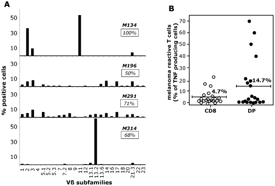
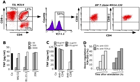
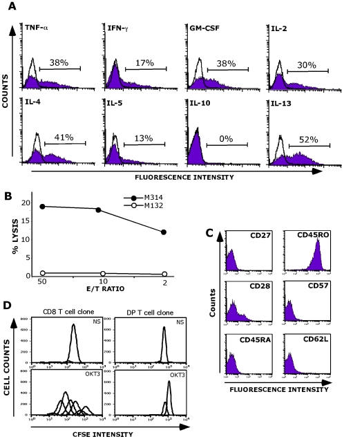
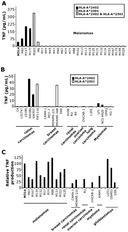
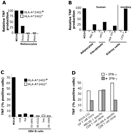
Methodology/principal findings: We analyzed the intratumoral cell infiltrate in solid metastasis (n = 6) and tumor invaded lymph nodes (n = 26) samples from melanomas patients by multiparametric cytometry. Here we documented for the first time significant increased frequency of DP T cells in about 60% of melanoma tumors compared to blood samples. Interestingly, a high proportion of these cells produced TNF-alpha in response to autologous melanoma cell lines. Besides, they are characterized by a unique cytokine profile corresponding to higher secretion of IL-13, IL-4 and IL-5 than simple positive T cells. In deep analysis, we derived a representative tumor-reactive DP T cell clone from a melanoma patient's invaded lymph node. This clone was restricted by HLA-A*2402 and recognized both autologous and allogeneic tumor cells of various origins as well as normal cells, suggesting that the target antigen was a ubiquitous self antigen. However, this DP T cell clone failed to kill HLA-A*2402 EBV-transformed B cells, probably due to the constitutive expression of immunoproteasome by these cells.

Conclusions/significance: In conclusion, we can postulate that, according to their broad tumor reactivity and to their original cytokine profile, the tumor associated DP T cells could participate in immune responses to tumors in vivo. Therefore, the presence of these cells and their role will be crucial to address in cancer patients, especially in the context of immunotherapies.


Conflict of interest statement

Competing Interests: The authors have declared that no competing interests exist.

Figures

Figure 1
Figure 1. Analysis of polyclonal DP T cells in melanoma: repertoire diversity and autologous-tumor reactivity.
A/ Repertoire diversity of DP T cells populations in tumors from four melanoma patients was assessed by labeling with 24 anti-Vβ mAbs. Insets indicate the percentage of the specific population characterized with this panel. B/ Percentage of TNF-producing cells in the SP CD8 and DP sub-populations in response to the autologous melanoma cell line. The autologous tumor cells lines were established from metastatic lymph nodes of 21 melanoma patients and were tested after two at four weeks of culture initiation. 106 ILNL and 2×10⁶ melanoma cells were incubated for 6 h in the presence of Brefeldin A, stained with CD4 and CD8 mAbs, fixed, and stained with anti-TNF Ab in a permeabilization buffer. 105 cells were then analyzed by flow cytometry.
Figure 2
Figure 2. DP T cell clone selection and characterization.
A/. Distribution of CD3+ T cells subsets based on CD4, CD8 in the M314 ILNL population. Lymphocytes were analyzed after in vitro expansion by three-color flow cytometry using antibodies specific for CD3, CD4 and CD8α or CD8β. Percentage of Vβ13.2 TCR expressing cells was examined on gated the 14% of DP T cells. The Vβ13.2 TCR expressing cells DP T cells were then sorted by FACS. B and C/ TNF secretion by the M314.132 DP T cell clone in response to the autologous melanoma cell line. 104 DP T cells were added to 3×10⁴ M314 melanoma cells in the presence or not of blocking antibodies directed against class I (W6/32), B/C/A24 (B1.23.2), class II (206) HLA and against CD4 and CD8 molecules at the indicated dilutions or concentrations. DP T cell clone reactivity was assessed by a TNF release assay. D/ Time course of CD3 (white) and TCR (black) down-regulation in M314.132 DP T cell clone stimulated with autologous melanoma cell line.
Figure 3
Figure 3. Functional properties of the M314.132 DP T cell clone.
A/ Cytokine production analysis. DP T cell clone was fixed, permeabilized and stained for cytokines following autologous melanoma stimulation. Data are expressed as mean % of intracellular cytokine secreting cells. Open histograms correspond to the analysis of cytokine production by unstimulated M314.132 DP T cell clone (negative control). B/Lysis of the M314 autologous melanoma cell line (closed circles) by M314.132 DP T cell clone. The M132 cell line was used as negative control target (open circles). 51Cr-labeled tumor cells were co-cultured with T cells at various E/T ratios. Chromium release in the supernantants was measured after a 4-h incubation period. C/ Phenotypic characterization of M314 DP T cell clone. D/ Proliferation capacity. CFSE-labeled T cell clones were stimulated with anti-CD3 (OKT3). The CD8 T cell clone used as positive control was obtained by limiting dilution of melanoma specific CD8 T cells. As negative control, T cell clones were maintained in the absence of any stimulation (Not Stimulated: NS).
Figure 4
Figure 4. Reactivity of M314.132 DP T cell clone against tumor cell lines.
A and B/ TNF secretion by the M314.132 DP T cell clone in response to tumor cell lines. 104 DP T cells were added to 3×10⁴ M314 melanoma cells (A) or other tumor cells (B). All tumor cell lines which are not recognized by the DP clone do not express the HLA-A*2402 and/or 2301 molecules. C/ TNF secretion by the M314.132 DP T cell clone to HLA-A*2402 transfected tumor cells. Melanoma (n = 10), breast carcinoma (n = 2), renal carcinoma (n = 1), ovarian carcinoma (n = 1), myeloma (n = 1), and glioblastoma (n = 3) cell lines were transiently transfected with 100ng of HLA-A*2402 plasmid with a lipofectamine reagent kit. 104 DP T cells were added to 3×10⁴ target cells, and the DP T cell clone reactivity was assessed by a TNF release assay. Results are expressed as relative reactivity to the indicated cells in comparison with TNF secretion (100%) induced by M314 autologous melanoma cells.
Figure 5
Figure 5. Reactivity of M314.132 DP T cell clone against normal cell lines.
A/ TNF secretion by the M314.132 DP T cell clone in response to melanocytes. 104 DP T cells were added to 3×10⁴ HLA-A*2402 positive or negative melanocytes, and the clone reactivity was assessed by a TNF release assy. B/ TNF secretion by the M314.132 DP T cell clone to HLA-A*2402 transfected (+) or non-transfected (−) normal cells of different origins and/or species. Results are expressed as relative reactivity to the indicated cells in comparison with TNF secretion (100%) induced by M314 autologous melanoma cells. C/ Lack of recognition of HLA-A*2402 EBV-B lymphocytes. DP T cell clone was fixed, permeabilized and stained for cytokines following stimulation with EBV-B cell lines expressing or not HLA-A*2402 molecules. Data are expressed as mean % of intracellular TNF-α secreting cells. D/ TNF response of DP T cell clone toward melanoma cells treated with or without IFN-γ. Melanoma cells were cultured in the presence or absence of 100U/ml rIFN-γ for 15 days. DP T cell clone and two CD8 T cell clones used as controls were fixed, permeabilized and stained for TNF following stimulation with untreated (white bars) or IFN-γ-treated (hatched bars) melanoma cells. Data are expressed as mean % of intracellular TNF-α secreting cells.

Similar articles

Cited by

References

    1. Dunn GP, Old LJ, Schreiber RD. The immunobiology of cancer immunosurveillance and immunoediting. Immunity. 2004;21(2):137–148. - PubMed
    1. Morgan RA, Dudley ME, Wunderlich JR, Hughes MS, Yang JC, et al. Cancer regression in patients after transfer of genetically engineered lymphocytes. Science. 2006;314(5796):126–129. - PMC - PubMed
    1. Rosenberg SA. Progress in human tumour immunology and immunotherapy. Nature. 2001;411(6835):380–384. - PubMed
    1. Appay V, Douek DC, Price DA. CD8+ T cell efficacy in vaccination and disease. Nat Med. 2008;14(6):623–628. - PubMed
    1. Khammari A, Labarriere N, Vignard V, Nguyen JM, Pandolfino MC, et al. Treatment of Metastatic Melanoma with Autologous Melan-A/Mart-1-Specific Cytotoxic T Lymphocyte Clones. J Invest Dermatol 2009 - PubMed

Publication types