. 2010 Feb;119(2):177-87.
doi: 10.1007/s00401-009-0633-x. Epub 2010 Jan 6.

Recombinant prion protein induces a new transmissible prion disease in wild-type animals

Affiliations

Recombinant prion protein induces a new transmissible prion disease in wild-type animals

Natallia Makarava et al. Acta Neuropathol. 2010 Feb.

Abstract

Prion disease is a neurodegenerative malady, which is believed to be transmitted via a prion protein in its abnormal conformation (PrP(Sc)). Previous studies have failed to demonstrate that prion disease could be induced in wild-type animals using recombinant prion protein (rPrP) produced in Escherichia coli. Here, we report that prion infectivity was generated in Syrian hamsters after inoculating full-length rPrP that had been converted into the cross-beta-sheet amyloid form and subjected to annealing. Serial transmission gave rise to a disease phenotype with highly unique clinical and neuropathological features. Among them were the deposition of large PrP(Sc) plaques in subpial and subependymal areas in brain and spinal cord, very minor lesioning of the hippocampus and cerebellum, and a very slow progression of disease after onset of clinical signs despite the accumulation of large amounts of PrP(Sc) in the brain. The length of the clinical duration is more typical of human and large animal prion diseases, than those of rodents. Our studies establish that transmissible prion disease can be induced in wild-type animals by inoculation of rPrP and introduce a valuable new model of prion diseases.


Figures

Fig. 1
Fig. 1
De novo generation of PrPSc in Syrian hamsters. a Western blotting of rPrP fibrils annealed in the presence of NBH or BSA. NBH-annealed fibrils showed a PK-resistant product of 16 kDa. Undigested samples were loaded at 1/20th the amount of the digested samples. b Western blotting of BHs from animals inoculated with NBH-annealed fibrils. Animal #36798 showed PK-resistant PrPSc with a characteristic band shift. c Western blotting of BHs from animals inoculated with BH from animal #36798. Without PK treatment, 1/10th of a sample was loaded. d Western blotting of BHs from animals inoculated with 10% BH from animal #36793. Undigested sample was loaded at 1/10th the amount of the digested samples. e Western blotting of BHs from animals inoculated with NBH-annealed fibrils. BHs were subjected to a single round of PMCA that consisted of 40-s sonication pulses applied every hour during a 48-h period, where each sample was its own substrate. Samples in a, b, e were treated with 50 μg/ml PK, and samples in c and d with 20 μg/ml PK. 3F4 antibody was used for a, ce and R1 antibody was used for b
Fig. 2
Fig. 2
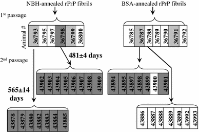
A flow chart outlining the serial passages of NBH- or BSA-annealed rPrP fibrils. Animal numbers are shown in the boxes. The animals that produced PrPSc detectible by Western blotting are in dark gray boxes, and the animals where PrPSc was detected after a single round of PMCA are in light gray boxes. Animal #36787 showed atypical, low molecular weight PK-resistant bands
Fig. 3
Fig. 3
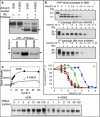
Biochemical characterization of the de novo generated prions. a BHs from the first (animal #36798) or second passage (animals #43902 and #43908) of NBH-annealed fibrils were treated with PK and PNGase and analyzed by Western blotting. Arrows indicate the center of the deglycosylated PrPSc from 263K-inoculated hamsters. BH #43902 was loaded at 1/4th of the amount of 263K BH. b Western blotting and the conformational stability profiles (iv) of the GdnHCl-induced transitions of the NBH-annealed fibrils (i), or PrPSc from first (ii) or second (iii) passage of NBH-annealed fibrils. NBH-annealed fibrils (blue circles), PrPSc from first passage (green square, animal #36798), second passage (yellow, orange, brown, or pink triangles—animals #43902, 43904, 43908, or 43909), or 263K-inoculated animals (red diamonds). Each BH was loaded onto the gel twice and the data were averaged. c The amplification kinetics for 263K or PrPSc from second passage of NBH-annealed fibrils (BH #43902) as monitored by Western blotting during a single round of PMCA that consisted of 100 cycles. Samples in a, b (ii and iii) were treated with 20 μg/ml PK; samples in c with 50 μg/ml PK; and samples in b (i) with 2 μg/ml PK
Fig. 4
Fig. 4
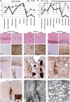
SSLOW strain displays unique neuropathological profile. a, b Lesion profile (a) and PrP immunopositivity score (b) in hamsters inoculated with SSLOW (2nd passage of NBH-annealed fibrils) or 263K. The lesion profile was obtained by averaging the scores for spongiform change, neuronal loss, and gliosis for three animals within each group. ce Comparison of spongiform changes in the hippocampus (c), thalamus (d), or cerebellum (e) stained with hematoxylin and eosin (upper panels) or anti-PrP 3F4 antibody (lower panels) in a representative hamsters inoculated with SSLOW (animal # 43902, left panels) or 263K (right panels). f Low-magnification (×20) overview of frontal cortex (enlarged at ×100 in right upper inset) and caudate-putamen (enlarged at ×100 in right lower inset) using immunostaining for disease-associated PrP. g, h High-magnification images (×400) of PrP immunoreactive plaques in the periventricular subependymal region (g), and synaptic and perineuronal PrP immunoreactivity in the frontal cortex (h). i Overview of PrP immunostaining (×50) of spinal cord demonstrates prominent plaque deposition (enlarged right lower inset) in the midline and in periaqueductal region. j, k Electron microscopy of a plaque from the periaqueductal region from the spinal cord (scale bar 10 μm in j or 0.4 μm in k)

Similar articles

Cited by

References

    1. Balch WE, Morimoto RI, Dillin A, Kelly JW. Adapting proteostasis for disease intervention. Science. 2008;319:916–919. doi: 10.1126/science.1141448. - DOI - PubMed
    1. Barria MA, Mukherjee A, Gonzalez-Romero D, Morales R, Soto C. De novo generation of infectious prions in vitro produces a new disease phenotype. PLOS Pathog. 2009;5:e1000421. doi: 10.1371/journal.ppat.1000421. - DOI - PMC - PubMed
    1. Baskakov IV, Breydo L. Converting the prion protein: what makes the protein infectious. Biochim Biophys Acta (Molecular Basis of Disease) 2007;1772:692–703. - PubMed
    1. Baskakov IV, Legname G, Baldwin MA, Prusiner SB, Cohen FE. Pathway complexity of prion protein assembly into amyloid. J Biol Chem. 2002;277:21140–21148. doi: 10.1074/jbc.M111402200. - DOI - PubMed
    1. Bocharova OV, Breydo L, Parfenov AS, Salnikov VV, Baskakov IV. In vitro conversion of full length mammalian prion protein produces amyloid form with physical property of PrPSc. J Mol Biol. 2005;346:645–659. doi: 10.1016/j.jmb.2004.11.068. - DOI - PubMed

Publication types