Review
. 2010 Apr;19(4):540-51.
doi: 10.1007/s00586-009-1251-y. Epub 2010 Jan 6.

Expanding role of 18F-fluoro-D-deoxyglucose PET and PET/CT in spinal infections

Affiliations
Review

Expanding role of 18F-fluoro-D-deoxyglucose PET and PET/CT in spinal infections

Filip Gemmel et al. Eur Spine J. 2010 Apr.

Abstract

(18)F-fluoro-D -deoxyglucose positron emission tomography ([(18)F]-FDG PET) is successfully employed as a molecular imaging technique in oncology, and has become a promising imaging modality in the field of infection. The non-invasive diagnosis of spinal infections (SI) has been a challenge for physicians for many years. Morphological imaging modalities such as conventional radiography, computed tomography (CT), and magnetic resonance imaging (MRI) are techniques frequently used in patients with SI. However, these methods are sometimes non-specific, and difficulties in differentiating infectious from degenerative end-plate abnormalities or postoperative changes can occur. Moreover, in contrast to CT and MRI, FDG uptake in PET is not hampered by metallic implant-associated artifacts. Conventional radionuclide imaging tests, such as bone scintigraphy, labeled leukocyte, and gallium scanning, suffer from relatively poor spatial resolution and lack sensitivity, specificity, or both. Initial data show that [(18)F]-FDG PET is an emerging imaging technique for diagnosing SI. [(18)F]-FDG PET appears to be especially helpful in those cases in which MRI cannot be performed or is non-diagnostic, and as an adjunct in patients in whom the diagnosis is inconclusive. The article reviews the currently available literature on [(18)F]-FDG PET and PET/CT in the diagnosis of SI.


Figures

Fig. 1
Fig. 1
Infected spinal hardware. A 53-year-old woman, who underwent spinal fusion with spinal implants 6 years earlier, was admitted to the hospital with an inflammatory syndrome of unknown origin. Posterior planar bone (a) and WBC images (c), demonstrate normal distribution of activity in the lumbar spine. Posterior planar gallium (b), and ciprofloxacin images (d), however, both demonstrate intense uptake in the lower lumbar spine and upper sacrum. Infected orthopedic hardware was surgically confirmed
Fig. 2
Fig. 2
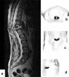
Chronic hematogenous spondylodiscitis of the thoracic spine. A 45-year-old man with multiple myeloma presents with severe back pain and prolonged fever in combination with increased inflammatory parameters. Sagittal T2-weighted MRI study (a) demonstrates abnormal signal at T11-12, but, nevertheless, the differential diagnosis with a vertebral compression fracture or even a spinal malignancy cannot reliably be made. Selected PET images (b transverse, c coronal, d sagittal view) demonstrate increased FDG uptake at the level of T11-12 vertebral disc, confirming a Staphylococcus aureus spondylodiscitis
Fig. 3
Fig. 3
Hematogenous spinal infection of the lumbar spine with secondary psoas abscess. A 60-year-old man, with known rheumatic disease (polychondritis/arthritis, episcleritis), treated with corticosteroids and methotrexate, presents with a grippal syndrome, malaise, chillings and fever up to 39°, and substantial weight loss. Laboratory results reveal increased inflammatory parameters (including a BSE of 56 and a CRP of 35 mg/dl). X-ray of the lumbar spine (a) revealed no pathological findings. Co-registered PET/enhanced CT (Biograph Siemens HiRez device) (selected sagittal CT, PET and fused PET/CT images, demonstrate high metabolic uptake at the L3–L4 level (b; dashed lines), extending in the soft prevertebral tissues
Fig. 4
Fig. 4
Same patient as in Fig. 3, high metabolic uptake localized in the right iliopsoas region (dashed lines, resp. a sagittal, b transverse, and c coronal CT, PET, and fused PET/CT images), indicating an occult spondylodiscitis complicated by a secondary psoas abscess; eventually the portal of entry was confirmed to be the urinary tract with a Staphylococcus aureus infection

References

    1. Adatepe MH, Powell OM, Isaacs GH, Nichols K, Cefola R. Hematogenous pyogenic vertebral osteomyelitis: diagnostic value of radionuclide bone imaging. J Nucl Med. 1986;27:1680–1685. - PubMed
    1. An HS, Seldomridge JA. Spinal infections: diagnostic tests and imaging studies. Clin Orthop Relat Res. 2006;444:27–33. doi: 10.1097/01.blo.0000203452.36522.97. - DOI - PubMed
    1. Aydinli U, Karaeminogullari O, Tiskaya K. Postoperative deep wound infection in instrumented spinal surgery. Acta Orthop Belg. 1999;65:182–187. - PubMed
    1. Boden SD, Davis DO, Dina TS, Sunner JL, Wiesel SW. Postoperative diskitis: distinguishing early MR imaging findings from normal postoperative disk space changes. Radiology. 1992;184:765–771. - PubMed
    1. Bredella MA, Essary B, Torriani M, Ouellette HA, Palmer WE. Use of FDG-PET in differentiating benign from malignant compression fractures. Skeletal Radiol. 2008;37:405–413. doi: 10.1007/s00256-008-0452-5. - DOI - PMC - PubMed

Substances