

Hepatocytes do not undergo epithelial-mesenchymal transition in liver fibrosis in mice
- PMID: 20052656
- PMCID: PMC2906231
- DOI: 10.1002/hep.23368
Hepatocytes do not undergo epithelial-mesenchymal transition in liver fibrosis in mice
Abstract
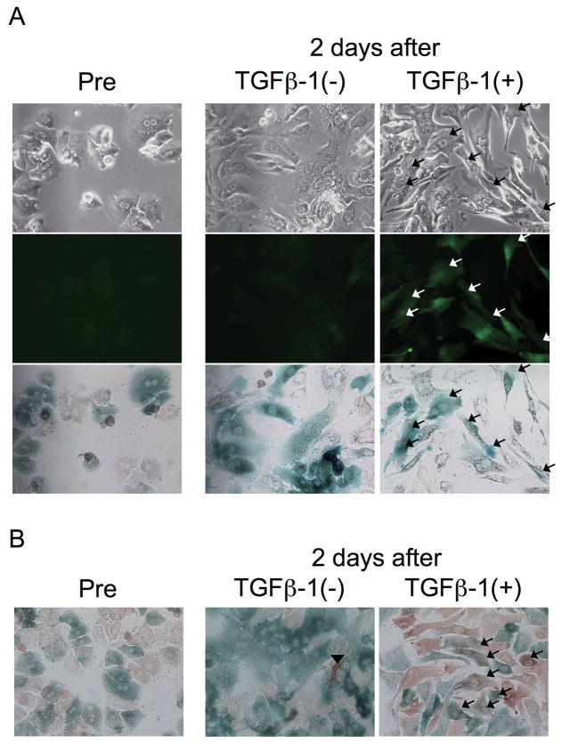
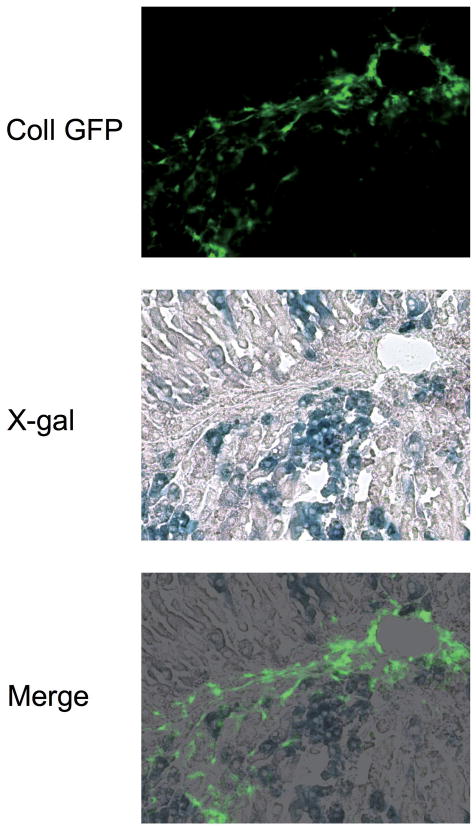
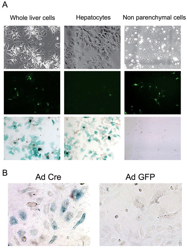
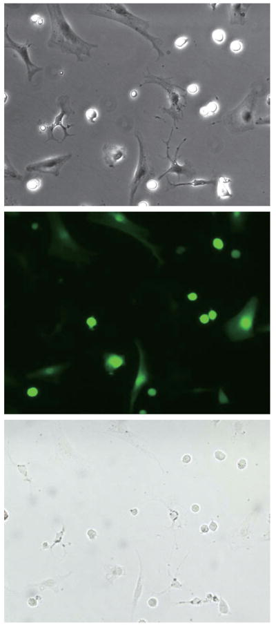
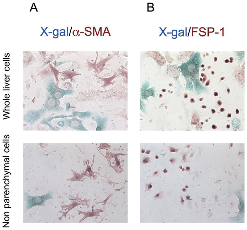
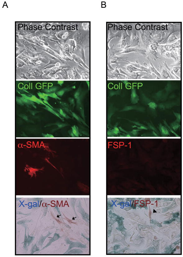
The origin of fibrogenic cells in liver fibrosis remains controversial. We assessed the emerging concept that hepatocytes contribute to production of extracellular matrix (ECM) in liver fibrosis through epithelial-mesenchymal transition (EMT). We bred triple transgenic mice expressing ROSA26 stop beta-galactosidase (beta-gal), albumin Cre, and collagen alpha1(I) green fluorescent protein (GFP), in which hepatocyte-derived cells are permanently labeled by beta-gal and type I collagen-expressing cells are labeled by GFP. We induced liver fibrosis by repetitive carbon tetrachloride (CCl(4)) injections. Liver sections and isolated cells were evaluated for GFP and beta-gal as well as expression of alpha-smooth muscle actin (alpha-SMA) and fibroblast-specific protein 1 (FSP-1). Upon stimulation with transforming growth factor beta-1, cultured hepatocytes isolated from untreated liver expressed both GFP and beta-gal with a fibroblast-like morphological change but lacked expression of other mesenchymal markers. Cells from CCl(4)-treated livers never showed double-positivity for GFP and beta-gal. All beta-gal-positive cells exhibited abundant cytoplasm, a typical morphology of hepatocytes, and expressed none of the mesenchymal markers including alpha-SMA, FSP-1, desmin, and vimentin. In liver sections of CCl(4)-treated mice, GFP-positive areas were coincident with fibrotic septa and never overlapped X-gal-positive areas.
Conclusion: Type I collagen-producing cells do not originate from hepatocytes. Hepatocytes in vivo neither acquire mesenchymal marker expression nor exhibit a morphological change clearly distinguishable from normal hepatocytes. Our results strongly challenge the concept that hepatocytes in vivo acquire a mesenchymal phenotype through EMT to produce the ECM in liver fibrosis.
Conflict of interest statement
No conflicts of interest exist.
Figures
Comment in
-
The epithelial-to-mesenchymal transition in liver fibrosis: here today, gone tomorrow?Hepatology. 2010 Mar;51(3):737-40. doi: 10.1002/hep.23529. Hepatology. 2010. PMID: 20198628 Free PMC article. No abstract available.
-
No contribution to liver fibrosis, but possible carcinogenesis?Hepatology. 2010 Apr;51(4):1468-9; author reply 1469. doi: 10.1002/hep.23604. Hepatology. 2010. PMID: 20373373 No abstract available.
References
-
- Beaussier M, Wendum D, Schiffer E, Dumont S, Rey C, Lienhart A, Housset C. Prominent contribution of portal mesenchymal cells to liver fibrosis in ischemic and obstructive cholestatic injuries. Lab Invest. 2007;87:292–303. - PubMed
-
- Kisseleva T, Uchinami H, Feirt N, Quintana-Bustamante O, Segovia JC, Schwabe RF, Brenner DA. Bone marrow-derived fibrocytes participate in pathogenesis of liver fibrosis. J Hepatol. 2006;45:429–438. - PubMed
Publication types
MeSH terms
Substances
Grants and funding
LinkOut - more resources
Full Text Sources
Other Literature Sources
Medical
Molecular Biology Databases
Research Materials