

Pelvic fractures after radiotherapy for cervical cancer: implications for survivors
- PMID: 20052724
- PMCID: PMC5084848
- DOI: 10.1002/cncr.24811
Pelvic fractures after radiotherapy for cervical cancer: implications for survivors
Abstract
Background: The incidence of pelvic fractures and associated risk factors was determined in women treated with curative-intent radiotherapy for cervical cancer.
Methods: The records of 516 women treated with curative-intent radiotherapy for cervical cancer between 2001 and 2006 at the University of Texas M. D. Anderson Cancer Center were reviewed. Among these, 300 patients had at least 1 post-treatment computed tomography scan or magnetic resonance imaging study available for review, and they comprised our study population. All imaging studies were re-reviewed by a single radiologist to evaluate for fractures.
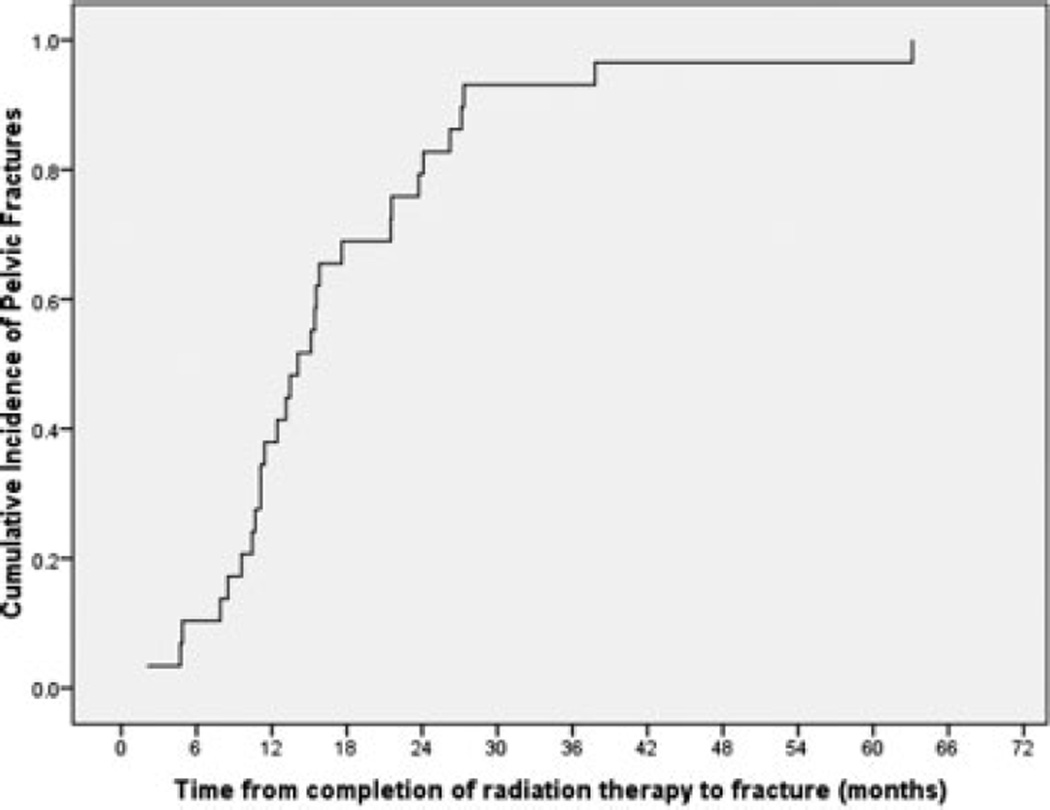
Results: Pelvic fractures were noted in 29 of 300 patients (9.7%). Fracture sites included sacrum (n = 24; 83%), sacrum and pubis (n = 3; 10%), iliac crest (n = 1; 3%), and sacrum and acetabulum (n = 1; 3%). Thirteen patients (45%) were symptomatic, with pain being the most common presenting symptom. The median time from the completion of radiotherapy to the detection of fractures on imaging studies was 14.1 months (range, 2.1-63.1 months), with 38% of patients diagnosed within 1 year and 83% diagnosed within 2 years of completing therapy. The median age of the patients at diagnosis was higher in the women who developed a fracture compared with the women who did not (56.5 years vs 46.7 years; P = .04). A higher number of women with a fracture were postmenopausal (62% vs 37%; P = .03). The median body mass index was lower in the women who had a fracture (26.0 kg/m2 vs 28.0 kg/m2; P = .03).
Conclusions: Pelvic fractures were detected in a substantial proportion of women after radiotherapy for cervical cancer. Bone mineral density screening and pharmacologic intervention should be considered in these women.
Copyright 2009 American Cancer Society.
Conflict of interest statement
DISCLOSURES The authors made no disclosures.
Figures
References
-
- Stehman FB, Bundy BN, DiSaia PJ, Keys HM, Larson JE, Fowler WC. Carcinoma of the cervix treated with radiation therapy. I. A multi-variate analysis of prognostic variables in the Gynecologic Oncology Group. Cancer. 1991;67:2776–2785. - PubMed
-
- Morris M, Eifel PJ, Lu J, et al. Pelvic radiation with concurrent chemotherapy compared with pelvic and para-aortic radiation for high-risk cervical cancer. N Engl J Med. 1999;340:1137–1143. - PubMed
-
- Rose PG, Bundy BN, Watkins EB, et al. Concurrent cisplatin-based radiotherapy and chemotherapy for locally advanced cervical cancer. N Engl J Med. 1999;340:1144–1153. - PubMed
-
- Keys HM, Bundy BN, Stehman FB, et al. Cisplatin, radiation, and adjuvant hysterectomy compared with radiation and adjuvant hysterectomy for bulky stage IB cervical carcinoma. N Engl J Med. 1999;340:1154–1161. - PubMed
-
- Rotman M, Pajak TF, Choi K, et al. Prophylactic extended-field irradiation of para-aortic lymph nodes in stages IIB and bulky IB and IIA cervical carcinomas. Ten-year treatment results of RTOG 79-20. JAMA. 1995;274:387–393. - PubMed
