. 2010 Apr;51(4):1420-9.
doi: 10.1002/hep.23427.

The anti-inflammatory effects of adiponectin are mediated via a heme oxygenase-1-dependent pathway in rat Kupffer cells

Affiliations

The anti-inflammatory effects of adiponectin are mediated via a heme oxygenase-1-dependent pathway in rat Kupffer cells

Palash Mandal et al. Hepatology. 2010 Apr.

Abstract

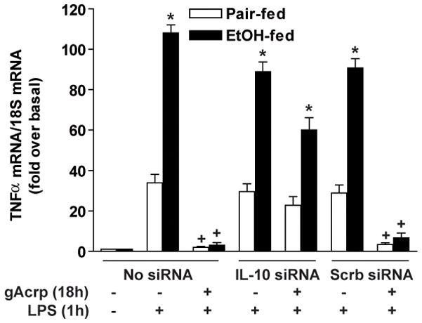
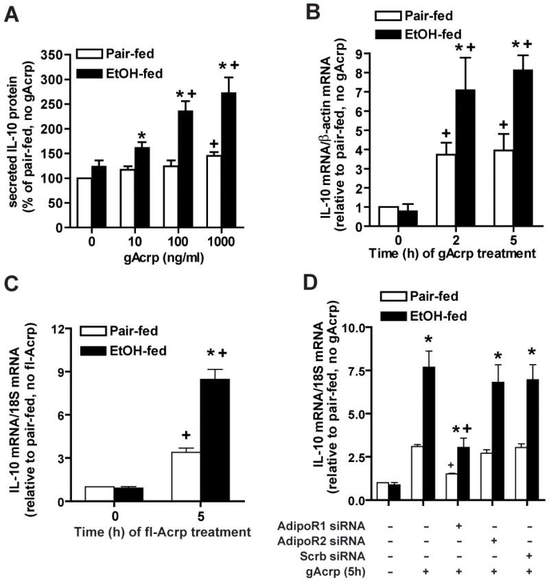
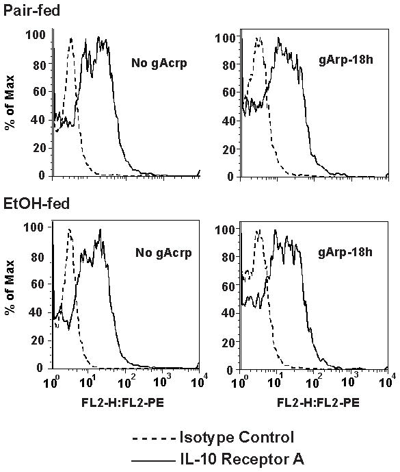
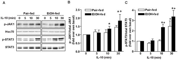
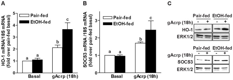
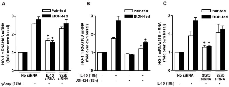
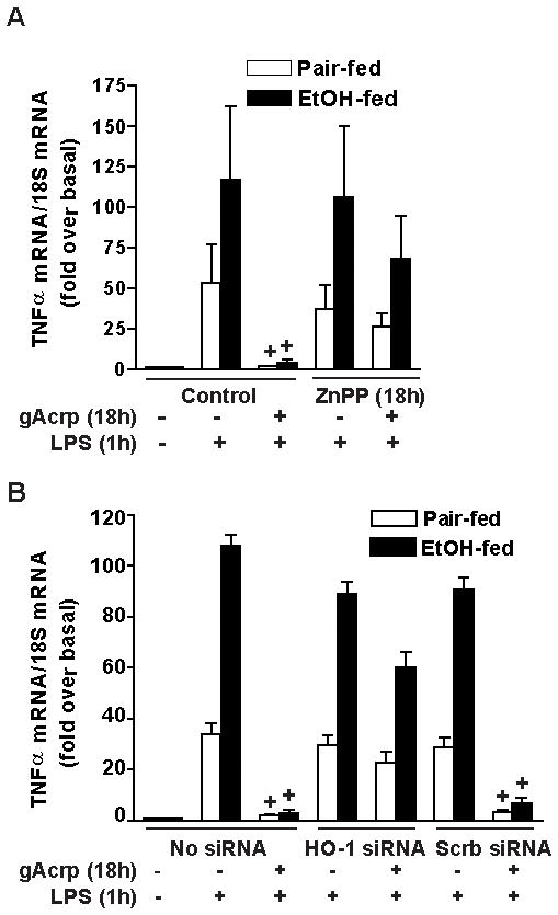
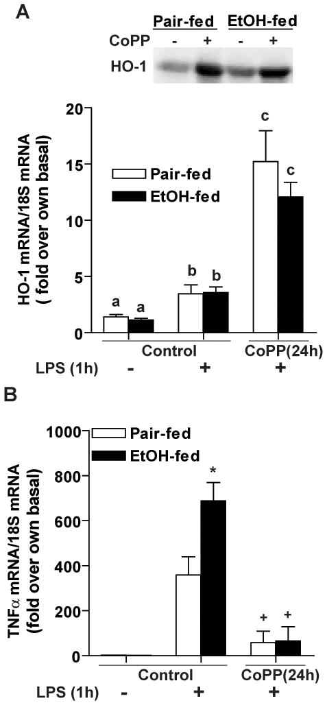
Altered expression and activity of immunomodulatory cytokines plays a major role in the pathogenesis of alcoholic liver disease. Chronic ethanol feeding increases the sensitivity of Kupffer cells, the resident hepatic macrophage, to lipopolysaccharide (LPS), leading to increased tumor necrosis factor alpha (TNF-alpha) expression. This sensitization is normalized by treatment of primary cultures of Kupffer cells with adiponectin, an anti-inflammatory adipokine. Here we tested the hypothesis that adiponectin-mediated suppression of LPS signaling in Kupffer cells is mediated via an interleukin-10 (IL-10)/heme oxygenase-1 (HO-1) pathway after chronic ethanol feeding. Knockdown of IL-10 expression in primary cultures of Kupffer cells with small interfering RNA (siRNA) prevented the inhibitory effect of globular adiponectin (gAcrp) on LPS-stimulated TNF-alpha expression. gAcrp increased IL-10 mRNA and protein expression, as well as expression of the IL-10 inducible gene, HO-1; expression was higher in Kupffer cells from ethanol-fed rats compared with pair-fed controls. Although IL-10 receptor surface expression on Kupffer cells was not affected by ethanol feeding, IL-10-mediated phosphorylation of STAT3 and expression of HO-1 was higher in Kupffer cells after ethanol feeding. Inhibition of HO-1 activity, either by treatment with the HO-1 inhibitor zinc protoporphyrin or by siRNA knockdown of HO-1, prevented the inhibitory effect of gAcrp on LPS-stimulated TNF-alpha expression in Kupffer cells. LPS-stimulated TNF-alpha expression in liver was increased in mice after chronic ethanol exposure. When mice were treated with cobalt protoporphyrin to induce HO-1 expression, ethanol-induced sensitivity to LPS was ameliorated.

Conclusion: gAcrp prevents LPS-stimulated TNF-alpha expression in Kupffer cells through the activation of the IL-10/STAT3/HO-1 pathway. Kupffer cells from ethanol-fed rats are highly sensitive to the anti-inflammatory effects of gAcrp; this sensitivity is associated with both increased expression and sensitivity to IL-10.


Figures

Figure 1
Figure 1. IL-10 siRNA prevents globular adiponectin (gAcrp)-induced suppression of TNF-α mRNA expression in LPS-stimulated Kupffer cells
Kupffer cells isolated from pair- and ethanol-fed rats were transfected or not with 2.0 μg of IL-10 siRNA or scrambled siRNA and then cultured with or without 1 μg/ml gAcrp for 18 h. Kupffer cells were then stimulated with 100 ng/ml of LPS for 60 min and TNF-α and 18S mRNA measured by qRT-PCR. In control experiments, siRNA knock-down of IL-10 decreased gAcrp-induced IL-10 mRNA expression after treatment with gAcrp for 5 h (see Supplementary Figure 1A). Knock-down efficiency did not differ between Kupffer cells from pair- and ethanol-fed rats. Values represent means ± SEM, n=4, *p<0.05 ethanol-fed compared to pair-fed, +p<0.05 compared to cells not treated with gAcrp.
Figure 2
Figure 2. Chronic ethanol feeding increases the sensitivity of Kupffer cells to adiponectin-stimulated IL-10 protein and mRNA expression
A) Kupffer cells isolated from pair- and ethanol-fed rats were cultured with 0–1000 ng/ml gAcrp for 18 h and IL-10 peptide secreted into the media measured by ELISA. Values represent means ± SEM, n=7, *p<0.05 ethanol-fed compared to pair-fed, +p<0.05 compared to cells not treated with gAcrp. B/C) Kupffer cells isolated from pair- and ethanol-fed rats were cultured overnight and then treated with 1 μg/ml gAcrp (B) or 1 μg/ml full-length adiponectin (C) for 0–5h and the quantity of IL-10, β-actin and 18S mRNA measured by qRT-PCR. Expression of IL-10 mRNA was normalized to β-actin or 18S and then expressed relative to expression in Kupffer cells from pair-fed rats not treated with gAcrp. Values represent means ± SEM, n=6–8 in B and 4 in C, *p<0.05 ethanol-fed compared to pair-fed, +p<0.05 compared to cells not treated with adiponectin. D) Kupffer cells were transfected or not with 2.0 μg of adiponectin R1 (AdipoR1), adiponectin R2 (AdipoR2) siRNA or scrambled siRNA and then cultured for 18 h. Kupffer cells were then stimulated or not with 1 μg/ml gAcrp for 5 h and IL-10 and 18S mRNA measured by qRT-PCR. siRNA knock-down decreased expression of AdipoR1 and AdipoR2 mRNA equally in Kupffer cells from pair- and ethanol-fed rats (see Supplementary Figure 1D/E). Values represent means ± SEM, n=4, *p<0.05 ethanol-fed compared to pair-fed, +p<0.05 compared to cells not treated with gAcrp.
Figure 3
Figure 3. IL-10 receptor A (IL-10RA) expression in Kupffer cells after chronic ethanol feeding
Kupffer cells isolated from pair- and ethanol-fed rats were treated with or without 1 μg/ml gAcrp for 18 h. and cell surface expression of IL-10RA (solid line), relative to isotype controls (dotted line), was measured by flow cytometry. Kupffer cells were harvested by gentle scraping and the cells collected by centrifugation at 300 × g for 10 minutes. The pellet was washed with PBS and resuspended in 100 μL PBS+ 0.1% sodium azide and then blocked with 1.0 μg anti-mouse CD32/CD16 Fcγ Block antibodies for 15 min at 4°C. Then the cells were stained with ~0.5 μg fluorochrome conjugated IL-10 receptor A (PE-conjugated IL-10 RA) or isotype control (PE-conjugated IgG1) diluted in PBS containing 0.1% sodium azide for 30 min. Cells were then washed twice with PBS and resuspended in 0.5 ml wash buffer (final concentration ~106 cells in 0.5 ml) and held on ice until flow cytometric measurements were performed on a FACScan flow cytometer (Becton Dickinson Immunocytometry systems, Mountain View, CA). The percentage of IL-10RA positive cells (50 ± 3 for pair-fed and 53 ± 3 for ethanol-fed) and the median fluorescence intensity (20 ± 8 for pair-fed and 26 ± 13) did not differ between Kupffer cells from pair- and ethanol-fed rats. Values represent means ± SEM, n=4. Traces shown are representative of four experiments.
Figure 4
Figure 4. Chronic ethanol feeding increases IL-10-stimulated phosphorylation of JAK1 and STAT3 in rat Kupffer cells
Kupffer cells isolated from pair-and ethanol-fed rats were cultured overnight and then stimulated with 10 ng/ml IL-10 for 0–30 min. Kupffer cells were then washed twice in ice-cold PBS and lysed at 4°C in 25 mM Tris-HCl, pH 7.6, 150 mM NaCl, 1% NP-40, 0.1% SDS containing 1 mM sodium orthovanadate, 10 mM NaF, 10 mM sodium pyrophosphate, 10 mM β-glycerophosphate, 1 mM PMSF, 10 μg/ml aprotinin and protease inhibitor (Complete-EDTA free). After 30 min, lysates were centrifuged at 16,000 × g for 15 min at 4°C. 20 μg of cellular extract was separated on 10% SDS-PAGE gels and then transferred to PVDF membranes for detection. Membranes were blocked in BSA and probed with antibodies against phospho-JAK1 and phospho-STAT-3. Membranes were then probed with antibodies to total JAK1 (data not shown), STAT3 and heat shock cognate protein 70 (hsc70), as a loading control. A) Representative images. B–C) Values represent means ± SEM, n=8–9, *p<0.05 ethanol-fed compared to pair-fed, +p<0.05 compared to cells (time 0) not treated with IL-10.
Figure 5
Figure 5. Chronic ethanol feeding increases gAcrp-mediated induction of HO-1 and SOCS-3 mRNA and HO-1 protein expression in rat Kupffer cells
Kupffer cells isolated from pair- and ethanol-fed rats were cultured with 1μg/ml gAcrp for 18h and quantity of A) heme-oxygenase-1 (HO-1) or B) suppressor of cytokine signaling-3 (SOCS-3) and 18S mRNA measured by qRT-PCR. HO-1 and SOCS-3 mRNA were normalized to 18S mRNA and values are expressed relative to Kupffer cells from pair-fed rats not treated with gAcrp. Values represent means ± SEM, n=4, values with different superscripts are significantly different from each other, p<0.05. C) Kupffer cells isolated from pair-and ethanol-fed rats were cultured with 1 μg/ml gAcrp for 18 h and HO-1 and SOCS-3 protein measured by Western blot. Images are representative of three independent experiments. gAcrp increased HO-1 protein by 1.66 ± 0.13 in pair-fed and 5.28 ± 1.73 (p<0.05 for ethanol-fed only, n=4).
Figure 6
Figure 6. Adiponectin induces HO-1 mRNA expression through an IL-10- and STAT3-dependent pathway
A) Kupffer cells isolated from pair- and ethanol-fed rats were transfected or not with 2.0 μg of IL-10 siRNA or scrambled siRNA and then cultured with or without 1 μg/ml gAcrp for 18 h. HO-1 and 18S mRNA were measured by qRT-PCR. HO-1 mRNA was normalized to 18S mRNA and values are expressed relative to Kupffer cells from pair-fed rats not treated with gAcrp. Values represent means ± SEM, n=3, +p<0.05 compared to gAcrp-treated cells not transfected with siRNA or transfected with scrambled siRNA. B) Kupffer cells isolated from pair- and ethanol-fed rats were pretreated with 10 μM JSI-124, an inhibitor of STAT3 signaling, or vehicle (DMSO) for 30 min and then cultured with or without 10 ng/ml IL-10 for 18h. HO-1 and 18S mRNA were measured by qRT-PCR. HO-1 mRNA was normalized to 18S mRNA and values are expressed relative to Kupffer cells from pair-fed rats not treated with IL-10. Values represent means ± SEM, n=3, +p<0.05 compared to IL-10-treated cells not treated with inhibitor. C) Kupffer cells isolated from pair- and ethanol-fed rats were transfected or not with 2.0 μg of STAT3 siRNA or scrambled siRNA and then cultured with or without 10 ng/ml IL-10 for 18 h. HO-1 and 18S mRNA were measured by qRT-PCR. HO-1 mRNA was normalized to 18S mRNA and values are expressed relative to Kupffer cells from pair-fed rats not treated with gAcrp. Values represent means ± SEM, n=3, +p<0.05 compared to gAcrp-treated cells not transfected with siRNA or transfected with scrambled siRNA.
Figure 7
Figure 7. HO-1 mediates the inhibitory effects of gAcrp on LPS-stimulated TNF-α expression
A) Kupffer cells isolated from pair- and ethanol-fed rats were cultured with or without 0.5 μM zinc protoporphyrin (ZnPP) in the presence or absence of 1μg/ml gAcrp for 18h. Kupffer cells were then stimulated with 100 ng/ml LPS for 1h and TNF-α and 18S mRNA measured by qRT-PCR. Values represent means ± SEM, n=4, +p<0.05 compared to control cells not treated with gAcrp. B) Kupffer cells isolated from pair- and ethanol-fed rats were transfected or not with 2.0 μg of HO-1 siRNA or scrambled siRNA and then cultured with or without 1 μg/ml gAcrp for 18 h. Kupffer cells were then stimulated with 100 ng/ml of LPS. TNF-α and 18S mRNA were measured by qRT-PCR. Values represent means ± SEM, n=5, +p<0.05 compared to cells within a treatment group not cultured with gAcrp.
Figure 8
Figure 8. Induction of HO-1 decreases LPS-stimulated TNF-α expression in vivo after chronic ethanol feeding to mice
A/B Mice were allowed free access to increasing concentrations of ethanol as part of a complete liquid diet to a maximum concentration of 32% of kcal over 25 days or pair-fed control diets (see Supplemental materials for detailed ethanol exposure protocol). Mice were then injected or not with 5 mg/kg cobalt protoporphyrin (CoPP) or vehicle (saline). After 24 h, mice were injected with 0.7 μg/kg LPS or saline. Expression of HO-1 (A) and TNF-α mRNA (B) was measured by qRT-PCR after 60 min and normalized to 18S mRNA. HO-1 protein was measured by Western blot (representative images are shown in the inset to panel A). Values represent means ± SEM, n=4–7 (A) values with difference superscripts are signficantly different from each other (B) *p<0.05 compared to pair-fed, +p<0.05 compared to LPS treated without treatment.

Similar articles

Cited by

References

    1. Vidali M, Stewart SF, Albano E. Interplay between oxidative stress and immunity in the progression of alcohol-mediated liver injury. Trends Mol Med. 2008;14:63–71. - PubMed
    1. O’Shea JJ, Murray PJ. Cytokine signaling modules in inflammatory responses. Immunity. 2008;28:477–487. - PMC - PubMed
    1. Le Moine O, Marchant A, De Groote D, Azar C, Goldman M, Deviere J. Role of defective monocyte interleukin-10 release in tumor necrosis factor-alpha overproduction in alcoholics cirrhosis. Hepatology. 1995;22:1436–1439. - PubMed
    1. Hill DB, D’Souza NB, Lee EY, Burikhanov R, Deaciuc IV, de Villiers WJ. A role for interleukin-10 in alcohol-induced liver sensitization to bacterial lipopolysaccharide. Alcohol Clin Exp Res. 2002;26:74–82. - PubMed
    1. Berg AH, Combs TP, Scherer PE. ACRP30/adiponectin: an adipokine regulating glucose and lipid metabolism. Trends Endocrinol Metab. 2002;13:84–89. - PubMed

Publication types

MeSH terms