. 2010 Jan;87(1):59-67.
doi: 10.1189/jlb.0409236.

The phenotype of murine wound macrophages

Affiliations

The phenotype of murine wound macrophages

Jean M Daley et al. J Leukoc Biol. 2010 Jan.

Abstract

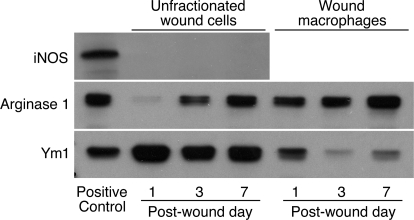
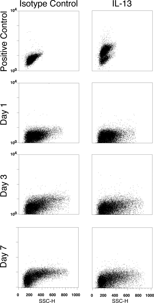
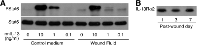
The phenotype of wound macrophages has not been studied by direct examination of these cells, yet macrophages recruited to sites of injury are described as alternatively activated macrophages, requiring IL-4 or IL-13 for phenotypic expression. This study characterized wound macrophage phenotype in the PVA sponge wound model in mice. Eighty-five percent of wound macrophages isolated 1 day after injury expressed Gr-1, but only 20% of those isolated at 7 days expressed this antigen. Macrophages from 1-, 3-, and 7-day wounds expressed markers of alternative activation,including mannose receptor, dectin-1, arginase 1,and Ym1, but did not contain iNOS. Day 1 wound macrophages produced more TNF-alpha, more IL-6, and less TGF-beta than Day 7 wound macrophages. Wound macrophages did not produce IL-10. The cytokines considered necessary for alternative activation of macrophages,IL-4 and IL-13, were not detected in the wound environment and were not produced by wound cells.Wound macrophages did not contain PStat6. Wound fluids inhibited IL-13-dependent phosphorylation of Stat6 and contained IL-13Ralpha2, a soluble decoy receptor for IL-13. The phenotype of wound macrophages was not altered in mice lacking IL-4Ralpha, which is required for Stat6-dependent signaling of IL-4 and IL-13.Wound macrophages exhibit a complex phenotype,which includes traits associated with alternative and classical activation and changes as the wound matures.The wound macrophage phenotype does not require IL-4 or IL-13.


Figures

Figure 1.
Figure 1.
Gr-1, mannose receptor (MR), and dectin-1 in wound macrophages. PVA sponges were inserted s.c. in B6D2F1 male mice. Wound cells were obtained 1, 3, or 7 days later. Leukocytes were isolated from blood. Cells were stained for Gr-1, mannose receptor, or dectin-1 (open histograms) or appropriate isotype control antibodies (shaded histograms). Additionally, wound cells were stained for CD68 and blood leukocytes for F4/80. Histograms show expression of antigens in gated macrophages (CD68high cells) or gated monocytes (F4/80+ cells). Results are representative of three experiments.
Figure 2.
Figure 2.
Arginase 1 and Ym1, but not iNOS, are present in wound macrophages. Wound cells were obtained 1, 3, or 7 days after PVA sponge insertion. Macrophages were isolated from the wound cell suspensions by negative selection. Cell lysates from unfractionated wound cells or isolated wound macrophages were size-fractionated and immunoblotted with antibodies for iNOS, arginase 1, and Ym1/2. Results are representative of two experiments.
Figure 3.
Figure 3.
Wound macrophages express mannose receptor and produce TNF-α. Wound cells were isolated 1 and 7 days after PVA sponge insertion, incubated in complete medium with Brefeldin A for 6 h, and stained with fluorochrome-conjugated antibodies for F4/80, TNF-α, and mannose receptor. The figure shows staining for TNF-α and mannose receptor in gated macrophages (F4/80+ cells). Results are representative of two experiments.
Figure 4.
Figure 4.
Wound cells do not produce IL-4. Wound cells were isolated at 1, 3, and 7 days after PVA sponge insertion, incubated in complete medium with Monensin for 6 h, and stained with fluorochrome-conjugated isotype control (left panels) or IL-4-specific (right panels) antibody. Positive control: BALB/c splenocytes cultured as described in Materials and Methods and stimulated with PMA and calcium ionomycin. Results are representative of two experiments. SSC-H, SSC-height.
Figure 5.
Figure 5.
Wound cells do not produce IL-13. Wound cells were isolated at 1, 3, and 7 days after PVA sponge insertion, incubated in complete medium with Brefeldin A for 6 h, and stained with fluorochrome-conjugated isotype control (left panels) or IL-13-specific (right panels) antibody. Positive control: BALB/c splenocytes cultured as described in Materials and Methods and stimulated with PMA and calcium ionomycin. Results are representative of two experiments.
Figure 6.
Figure 6.
Wound macrophages express IL-4Rα but do not contain PStat6. Wound cells were stained for IL-4Rα (open histograms) or with isotype control antibody (shaded histograms) and for CD68. Histograms show staining for IL-4Rα in gated macrophages (CD68high cells). For PStat6 staining, wound cells were incubated or not with IL-4 (10 ng/mL) for 15 min and then fixed and stained for PStat6 (open histograms) or with isotype control antibody (shaded histograms) and with Gr-1 and F4/80. Histograms show staining for PStat6 in gated macrophages (F4/80+, Gr-1low/int cells). Results are representative of two experiments.
Figure 7.
Figure 7.
Wound fluids inhibit rmIL-13-induced phosphorylation of Stat6 and contain IL-13Rα2. (A) J774A.1 cells were treated with rmIL-13, diluted in control media or pooled Day 1 wound fluids. Cells lysates were size-fractionated and immunoblotted with antibodies to PStat6 and Stat6. Identical results were obtained when rmIL-13 was added to Day 7 wound fluids (not shown). (B) Wound fluids obtained at 1, 3, and 7 days after PVA sponge insertion were size-fractionated and immunoblotted with antibodies to IL-13Rα2. Results are representative of two experiments.
Figure 8.
Figure 8.
Mannose receptor and dectin-1 in wound macrophages are independent of IL-4Rα. Wound cells were obtained from wild-type or IL-4Rα knockout (IL-4rα −/−) mice 7 days after PVA sponge insertion. Cells were stained for mannose receptor or dectin-1 (open histograms) or the appropriate isotype control antibodies (shaded histograms) and for CD68. Histograms show expression of the antigens in gated macrophages (CD68high cells).
Figure 9.
Figure 9.
iNOS, arginase I, and Ym1 in wound macrophages are independent of IL-4Rα. Wound cells were obtained from wild-type (W.T.) or IL-4Rα knockout mice 7 days after PVA sponge insertion. Macrophages were isolated by negative selection. Cell lysates from unfractionated wound cells or isolated wound macrophages were size-fractionated and immunoblotted with antibodies for iNOS, Arginase 1, and Ym1/2.

Comment in

Similar articles

Cited by

References

    1. Kodelja V, Muller C, Tenorio S, Schebesch C, Orfanos C E, Goerdt S. Differences in angiogenic potential of classically vs alternatively activated macrophages. Immunobiology. 1997;197:478–493. - PubMed
    1. Gordon S. Alternative activation of macrophages. Nat Rev Immunol. 2003;3:23–35. - PubMed
    1. Gordon S, Taylor P R. Monocyte and macrophage heterogeneity. Nat Rev Immunol. 2005;5:953–964. - PubMed
    1. Martinez F O, Helming L, Gordon S. Alternative activation of macrophages: an immunologic functional perspective. Annu Rev Immunol. 2009;27:451–483. - PubMed
    1. Raes G, De Baetselier P, Noel W, Beschin A, Brombacher F, Hassanzadeh Gh G. Differential expression of FIZZ1 and Ym1 in alternatively versus classically activated macrophages. J Leukoc Biol. 2002;71:597–602. - PubMed

Publication types

MeSH terms