. 2009:9:1-13.
doi: 10.1673/031.009.5501.

Growth and differentiation of the larval mosquito midgut

Affiliations

Growth and differentiation of the larval mosquito midgut

Kathryn Ray et al. J Insect Sci. 2009.

Abstract

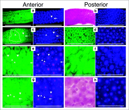
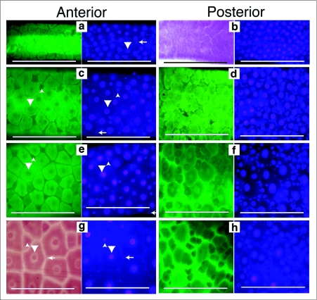
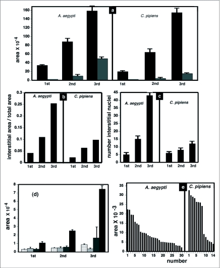
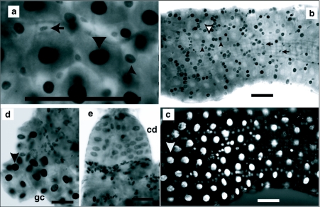
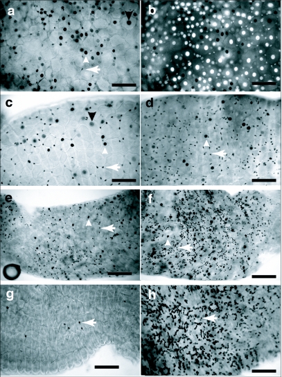
Factors affecting larval growth and nutrition have consequences on adult fecundity. Since the mosquito larval midgut is the primary organ of digestion and nutrient absorption, factors that affect the growth and development of the midgut may have potential consequences on the reproductive potential of the adult. To gain a better understanding of mosquito midgut development the growth and metamorphic remodeling of the Aedes aegypti L. and Culex pipiens L. (Diptera: Culicidae) midguts were investigated. Cytological evidence was obtained suggesting that, in both the anterior and posterior Ae. aegypti larval midgut, diploid regenerative cells give rise to new endoreplicating cells that significantly contribute to the growth and metabolism of the midgut. This hypothesis was supported by BrdU incorporation studies showing that diploid cells, as well as large and small endoreplicating cells, synthesize DNA during the 2(nd), 3(rd) and 4(th) instars. Cytological studies of the Cx. pipiens larval midgut suggest that anterior midgut growth in this species is primarily by cell enlargement. To study metamorphic remodeling of the midgut, DNA synthesis in Ae. aegypti 4(th) instar midguts was followed by using 5-bromo-2-deoxyuridine (BrdU) incorporation. During the 24 hr period after the last larval-larval molt both endoreplicating and diploid cells incorporate BrdU. After the critical weight is achieved, endoreplicating cell BrdU incorporation gradually ceases while diploid cells continue to replicate. The period of maximum diploid cell incorporation correlated with the period of maximum ecdysone titer.


Figures

Figure 1.
Figure 1.
Growth and development of the Aedes aegypti larval midgut. Midguts from 1st through 4th instars were dissected, fixed and stained with Hematoxylin and DAPI. First through 3rd instar midguts were dissected approximately 12 hours after the molt. Fourth instar midguts were dissected 24 hours after the molt. The left side image of each pair was taken using autofluorescence (green) or bright field (red), to emphasize the boundaries of the cells. The right side image is the same field of view showing DAPI stained nuclei. All images were taken at the same magnification and the scale bar represents 200 µm. Large nuclei are indicated by downward pointing arrow heads. Intermediate sized nuclei are indicated by upward pointing arrow heads. Small oval nuclei are indicated by left pointing arrows, (a) 1st instar anterior midgut had uniformly spaced large nuclei with interspersed small nuclei, (b) 1st instar posterior midgut had uniformly spaced large nuclei with interspersed small nuclei, (c) 2nd instar anterior midgut had uniformly spaced large nuclei and more interspersed smaller and intermediate sized nuclei, (d) 2nd instar posterior midgut had uniformly spaced large nuclei and numerous interspersed smaller and intermediate sized nuclei, (e) 3rd instar anterior midgut had uniformly spaced large nuclei that were surrounded by numerous intermediate and small sized nuclei, (f) 3rd instar posterior midgut had uniformly spaced large nuclei and many intermediate and small sized nuclei surrounding the large nuclei, (g) 4th instar anterior midgut had large and intermediate sized nuclei surrounded by small elongate nuclei, (h) 4th instar posterior midgut also had large and intermediate sized nuclei surrounded by elongate small nuclei.
Figure 2.
Figure 2.
Growth and development of the Culex pipiens larval midgut. Midguts from 1st through 4th instars were dissected, fixed and stained with Hematoxylin and DAPI. First through 3rd instar midguts were dissected approximately 12 hours after the molt. Fourth instar midguts were dissected 24 hours after the molt. Each subfigure has a left side image, taken using autofluorescence (green) or bright field (red), to emphasize the boundaries of the cells. The right side image is the same field of view to show DAPI stained nuclei. Large nuclei are indicated by downward pointing arrow heads. Intermediate sized nuclei are indicated by upward pointing arrow heads. Small oval nuclei are indicated by left pointing arrows. All images are taken at the same magnification and the scale bar represents 200 µm. (a) 1st instar anterior midgut have evenly spaced large nuclei with a few small nuclei between them, (b) 1st instar posterior midgut has evenly spaced large nuclei with more small nuclei between them, (c) 2nd instar anterior midgut has evenly spaced large nuclei and a few intermediate sized nuclei between them, (d) 2nd instar posterior midgut has evenly spaced large nuclei with many small and intermediate sized nuclei between them, (e) 3rd instar anterior midgut has evenly spaced large nuclei with a few intermediate sized nuclei between them, (f) 3rd instar posterior midgut has evenly spaced large nuclei with many intermediate and small sized nuclei between them, (g) 4th instar anterior midgut has evenly spaced large nuclei, a few intermediate sized and small nuclei between them, (h) 4th instar posterior midgut has many small and intermediate sized nuclei surrounding the large nuclei.
Figure 3.
Figure 3.
Measurements of Aedes aegypti and Culex pipiens midgut growth. Bright field and fluorescence images of midguts were used to measure growth in 1st through 3rd instar midguts that were dissected 12 hours after the molt. The areas of anterior midgut regions bounded by 10 adjacent cells with large nuclei were measured (black bars). An example is shown in Figure 1c. The areas of interstitial small and intermediate sized cells within the 10 cell regions were measured (grey bars). The number of interstitial small and intermediate sized nuclei within the 10 cell regions were counted. Areas are measured in pixels using ImageJ software, (a) Growth of anterior midgut from 1st to 3rd instars. Ten cell area (black bars); interstitial small and intermediate sized cell area (grey bars), (b) Fraction of anterior midgut growth due to interstitial small and intermediate sized cells, (c) Number of interstitial small and intermediate cell nuclei, (d) Sizes of nuclei in Ae. aegypti 1st through 3rd instars. Nuclei identified by their chain-like arrangement and circular shape (stippled bars). Small oval nuclei interspersed between larger nuclei (crosshatched bars). Intermediate sized nuclei, round in shape, and interspersed between largest nuclei (grey bars). Largest nuclei surrounded by small and intermediate sized nuclei (black bars), (e) Distribution of cell sizes in anterior midguts of Ae. aegypti and Cx. pipiens 4th instars. Squares with the dimensions of 0.3mm long by 0.3mm wide were marked in autofluorescence images of Ae. aegypti and Cx. pipiens 24 hour postmolt 4th instar anterior midguts and the sizes of the cells within these squares were measured.
Figure 4.
Figure 4.
Endoreplicating and diploid cells of 2nd and 3rd instars incorporate BrdU. Aedes aegypti 1st through 3rd instars were exposed to 100µg/ml BrdU for 24h. BrdU labeled large nuclei are indicated by solid downward pointing arrow head. Large nuclei not labeled with BrdU are indicated by open downward pointing arrowhead. Intermediate sized nuclei are indicated by upward pointing arrow head. Small oval nuclei are indicated by left pointing arrow. Scale bar represents 100 µm. (a) Anterior midgut of 2nd instar showing small oval, intermediate and large sized nuclei were BrdU labeled, (b) Posterior midgut of 3rd instar showing small oval and intermediate sized nuclei were labeled by BrdU. Large nuclei were not labeled, (c) Same field of view as presented in Figure 4b illuminated to reveal DAPI stained nuclei. Large nuclei are brightly fluorescent but unlabeled by BrdU. (d) Gastric caecum (gc) of 3rd instar showing labeled large nuclei and labeled diploid nuclei at the base of the gastric ceacum. (e) Cardia (cd) of 3rd instar showing labeled large nuclei and small nuclei at the base of the cardia.
Figure 5.
Figure 5.
BrdU incorporation during midgut metamorphosis in Aedes aegypti 4th instars exposed to 100 µg/ml BrdU for the time intervals indicated below. Staining was performed as described in the Methods. BrdU labeled large nuclei are indicated by a downward pointing arrow head. Intermediate sized nuclei are indicated by an upward pointing arrow head. Small oval nuclei are indicated by left pointing arrow. All images were taken at the same magnification and scale bar represents 100 µm. (a) Anterior midgut of 4th instar exposed to BrdU from the time of molt to 14 hours after the molt, showing large, intermediate sized and small oval nuclei incorporated BrdU. (b) Same field as in Figure 4a but illuminated to detect DAPI staining. Most large nuclei were brightly fluorescent but not BrdU labeled, (c) Anterior midgut of 4th instar exposed to BrdU from 14 to 24 hours after the molt. Large, intermediate sized and small oval nuclei incorporated BrdU. (d) Posterior midgut of 4th instar exposed to BrdU from 14 to 24 hours after the molt. Incorporation by small oval nuclei greater than in Figure 5c. A few intermediate sized nuclei incorporated BrdU. (e) Anterior midgut of 4th instar exposed to BrdU from 24 to 36 hours after the molt. BrdU incorporation was predominately in small oval nuclei, (f) Posterior midgut of 4th instar exposed to BrdU from 24 to 36 hours after the molt. Labeled small oval nuclei were more numerous but a few intermediate sized cells were labeled, (g) Anterior midgut of 4th instar exposed to BrdU from 38 to 48 hours after the molt showed very little incorporation, (h) Posterior midgut of 4th instar exposed to BrdU from 38 to 48 hours after the molt showed many labeled diploid nuclei and no labeled large nuclei.

Similar articles

Cited by

References

    1. Baldwin KM, Hakim RS. Growth and differentiation of the larval midgut epithelium during molting in the moth, Manduca sexta. . Tissue and Cell. 1991;23:411–422. - PubMed
    1. Berger CA. Cytology of metamorphosis in the Culininae. Nature. 1938;141:834–835.
    1. Bernick EP, Moffett SB, Moffett DF. Organization, ultrastructure, and development of midgut visceral muscle in larval Aedes aegypti. . Tissue and Cell. 2007;39:277–292. - PMC - PubMed
    1. Briegel H. Fecundity, metabolism, and body size in Anopheles (Diptera: Culicidae), vectors of malaria. Journal of Medical Entomology. 1990;27:839–850. - PubMed
    1. Briegel H. Physiological basis of mosquito ecology. Journal of Vector Ecology. 2003;28:1–11. - PubMed

Publication types