Lack of demonstrable memory T cell responses in humans who have spontaneously recovered from leptospirosis in the Peruvian Amazon
- PMID: 20053135
- PMCID: PMC2877911
- DOI: 10.1086/650300
Lack of demonstrable memory T cell responses in humans who have spontaneously recovered from leptospirosis in the Peruvian Amazon
Abstract
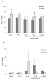
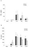
BACKGROUND. We tested the hypothesis that patients who have recovered from leptospirosis have peripheral blood memory T cells that are specific for Leptospira or Leptospira protein antigens. METHODS. Peripheral blood mononuclear cells (PBMCs) were obtained from patients who had recovered from leptospirosis, as well as from control individuals. PBMCs were assessed for in vitro proliferation, phenotyping, and cytokine production after stimulation with different strains of Leptospira, recombinant LipL32, or overlapping synthetic peptides of different outer membrane proteins. RESULTS. PBMCs from both control subjects and patients produced significant proliferative responses to all Leptospira strains. Proliferation from control PBMCs was significantly greater than responses produced by patient PBMCs. Select strains of Leptospira expanded both T cell receptor (TCR) alphabeta and TCRgammadelta T cells in both control subject and patient PBMCs. Patient and control subject PBMCs produced equivalent levels of tumor necrosis factor alpha and interferon gamma, but patient PBMCs produced significantly less interleukin 10 than did control subject PBMCs after stimulation by different strains of Leptospira. PBMCs from patients failed to respond to recombinant LipL32 or to any of the Leptospira peptides. CONCLUSION. Leptospira induced significant proliferative responses, TCRgammadelta T cell expansion, and cytokine production in both control subject and patient PBMCs. Patient PBMCs failed to recognize Leptospira protein antigens. Leptospirosis does not seem to generate memory T cells that can be activated by in vitro stimulation.
Conflict of interest statement
None of the authors has any conflict of interest in the work presented.
Figures


Similar articles
-
Bovine Immune Response to Vaccination and Infection with Leptospira borgpetersenii Serovar Hardjo.mSphere. 2021 Mar 24;6(2):e00988-20. doi: 10.1128/mSphere.00988-20. mSphere. 2021. PMID: 33762318 Free PMC article.
-
Leptospira interrogans activation of human peripheral blood mononuclear cells: preferential expansion of TCR gamma delta+ T cells vs TCR alpha beta+ T cells.J Immunol. 2003 Aug 1;171(3):1447-55. doi: 10.4049/jimmunol.171.3.1447. J Immunol. 2003. PMID: 12874237
-
Immunogenicity of a DNA and Recombinant Protein Vaccine Combining LipL32 and Loa22 for Leptospirosis Using Chitosan as a Delivery System.J Microbiol Biotechnol. 2015 Apr;25(4):526-36. doi: 10.4014/jmb.1408.08007. J Microbiol Biotechnol. 2015. PMID: 25348693
-
Leptospira and leptospirosis in China.Curr Opin Infect Dis. 2014 Oct;27(5):432-6. doi: 10.1097/QCO.0000000000000097. Curr Opin Infect Dis. 2014. PMID: 25061933 Review.
-
Leptospirosis: a Toll road from B lymphocytes.Chang Gung Med J. 2010 Nov-Dec;33(6):591-601. Chang Gung Med J. 2010. PMID: 21199604 Review.
Cited by
-
Biannual and Quarterly Comparison Analysis of Agglutinating Antibody Kinetics on a Subcohort of Individuals Exposed to Leptospira interrogans in Salvador, Brazil.Front Med (Lausanne). 2022 Apr 14;9:862378. doi: 10.3389/fmed.2022.862378. eCollection 2022. Front Med (Lausanne). 2022. PMID: 35492362 Free PMC article.
-
Estimating leptospirosis incidence using hospital-based surveillance and a population-based health care utilization survey in Tanzania.PLoS Negl Trop Dis. 2013 Dec 5;7(12):e2589. doi: 10.1371/journal.pntd.0002589. eCollection 2013. PLoS Negl Trop Dis. 2013. PMID: 24340122 Free PMC article.
-
LigA formulated in AS04 or Montanide ISA720VG induced superior immune response compared to alum, which correlated to protective efficacy in a hamster model of leptospirosis.Front Immunol. 2022 Oct 10;13:985802. doi: 10.3389/fimmu.2022.985802. eCollection 2022. Front Immunol. 2022. PMID: 36300125 Free PMC article.
-
Association Between Severity of Leptospirosis and Subsequent Major Autoimmune Diseases: A Nationwide Observational Cohort Study.Front Immunol. 2021 Sep 10;12:721752. doi: 10.3389/fimmu.2021.721752. eCollection 2021. Front Immunol. 2021. PMID: 34566978 Free PMC article.
-
Severe Leptospirosis Features in the Spleen Indicate Cellular Immunosuppression Similar to That Found in Septic Shock.Front Immunol. 2019 Apr 30;10:920. doi: 10.3389/fimmu.2019.00920. eCollection 2019. Front Immunol. 2019. PMID: 31114579 Free PMC article.
References
-
- Vinetz JM. Leptospirosis. Curr. Opin. Infect. Dis. 2001;14:527. - PubMed
-
- World Health Organization. Leptospirosis worldwide, 1999. Wkly. Epidemiol. Rec. 1999. p. 237. - PubMed
-
- Ashford DA, Kaiser RM, Spiegel RA, et al. Asymptomatic infection and risk factors for leptospirosis in Nicaragua. Am. J. Trop. Med. Hyg. 2000;63:249. - PubMed
-
- Berman SJ, Tsai CC, Holmes K, Fresh JW, Watten RH. Sporadic anicteric leptospirosis in South Vietnam. A study in 150 patients. Ann. Intern. Med. 1973;79:167. - PubMed

