

Transgenic mice enriched in omega-3 fatty acids are more susceptible to pulmonary tuberculosis: impaired resistance to tuberculosis in fat-1 mice
- PMID: 20053136
- PMCID: PMC4421876
- DOI: 10.1086/650344
Transgenic mice enriched in omega-3 fatty acids are more susceptible to pulmonary tuberculosis: impaired resistance to tuberculosis in fat-1 mice
Abstract
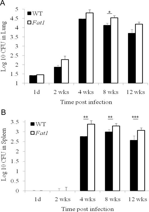
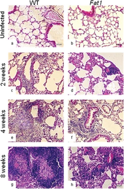
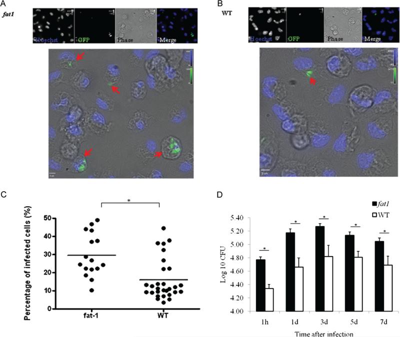
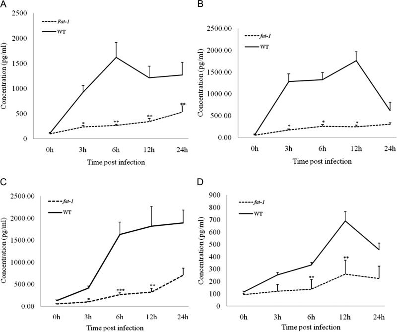
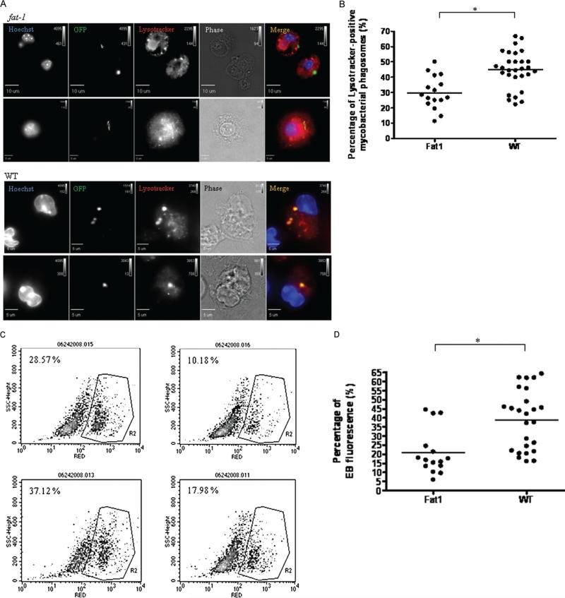
BACKGROUND. Besides their health benefits, dietary omega-3 fatty acids (n-3 PUFAs) can impair host resistance to intracellular pathogens. Previously, we and others have showed that n-3 PUFA-treated macrophages poorly control Mycobacterium tuberculosis infection in vitro. METHODS. Wild-type and fat-1 transgenic mice were infected with virulent H37Rv M. tuberculosis via the aerosol route. We evaluated bacteriological and histopathological changes in lungs, as well as differences in activation and antimycobacterial capacity in primary macrophages ex vivo. RESULTS. fat-1 mice were more susceptible to tuberculosis, as demonstrated by higher bacterial loads and less robust inflammatory responses in lungs. Macrophages obtained from fat-1 mice were more readily infected with M. tuberculosis in vitro, compared with wild-type macrophages. This impaired bacterial control in cells from fat-1 mice correlated with reduced proinflammatory cytokine secretion, impaired oxidative metabolism, and diminished M. tuberculosis-lysotracker colocalization within phagosomes. CONCLUSIONS. We showed that endogenous production of n-3 PUFAs in fat-1 mice increases their susceptibility to tuberculosis, which could be explained in part by diminished activation and antimycobacterial responses in cells from fat-1 mice. These data suggest that n-3 PUFA-supplemented diets might have a detrimental effect on immunity to M. tuberculosis and raise concerns regarding the safety of omega-3 dietary supplementation in humans.
Figures
Similar articles
-
Incorporation of a dietary omega 3 fatty acid impairs murine macrophage responses to Mycobacterium tuberculosis.PLoS One. 2010 May 28;5(5):e10878. doi: 10.1371/journal.pone.0010878. PLoS One. 2010. PMID: 20526363 Free PMC article.
-
Inhibition of Pancreatic Carcinoma Growth Through Enhancing ω-3 Epoxy Polyunsaturated Fatty Acid Profile by Inhibition of Soluble Epoxide Hydrolase.Anticancer Res. 2019 Jul;39(7):3651-3660. doi: 10.21873/anticanres.13513. Anticancer Res. 2019. PMID: 31262891 Free PMC article.
-
Endogenous ω-3 fatty acids in Fat-1 mice attenuated depression-like behaviors, spatial memory impairment and relevant changes induced by olfactory bulbectomy.Prostaglandins Leukot Essent Fatty Acids. 2021 Aug;171:102313. doi: 10.1016/j.plefa.2021.102313. Epub 2021 Jun 24. Prostaglandins Leukot Essent Fatty Acids. 2021. PMID: 34246927
-
From fat to fat-1: a tale of omega-3 fatty acids.J Membr Biol. 2005 Jul;206(2):165-72. doi: 10.1007/s00232-005-0790-3. J Membr Biol. 2005. PMID: 16456726 Review. No abstract available.
-
n-3 polyunsaturated fatty acids and HER2-positive breast cancer: interest of the fat-1 transgenic mouse model over conventional dietary supplementation.Biochimie. 2014 Jan;96:22-7. doi: 10.1016/j.biochi.2013.08.021. Epub 2013 Sep 6. Biochimie. 2014. PMID: 24012777 Review.
Cited by
-
Dietary long-chain omega 3 fatty acids modify sphingolipid metabolism to facilitate airway hyperreactivity.Sci Rep. 2022 Nov 17;12(1):19735. doi: 10.1038/s41598-022-21083-w. Sci Rep. 2022. PMID: 36396956 Free PMC article.
-
Incorporation of a dietary omega 3 fatty acid impairs murine macrophage responses to Mycobacterium tuberculosis.PLoS One. 2010 May 28;5(5):e10878. doi: 10.1371/journal.pone.0010878. PLoS One. 2010. PMID: 20526363 Free PMC article.
-
Omega-3 Polyunsaturated Fatty Acids Prevent Toxoplasma gondii Infection by Inducing Autophagy via AMPK Activation.Nutrients. 2019 Sep 6;11(9):2137. doi: 10.3390/nu11092137. Nutrients. 2019. PMID: 31500218 Free PMC article.
-
n-3 Polyunsaturated Fatty Acids Impede the TCR Mobility and the TCR-pMHC Interaction of Anti-Viral CD8+ T Cells.Viruses. 2020 Jun 12;12(6):639. doi: 10.3390/v12060639. Viruses. 2020. PMID: 32545480 Free PMC article.
-
Narrative Review of n-3 Polyunsaturated Fatty Acid Supplementation upon Immune Functions, Resolution Molecules and Lipid Peroxidation.Nutrients. 2021 Feb 18;13(2):662. doi: 10.3390/nu13020662. Nutrients. 2021. PMID: 33670710 Free PMC article. Review.
References
-
- Anes E, Kuhnel MP, Bos E, Moniz-Pereira J, Habermann A, Griffiths G. Selected lipids activate phagosome actin assembly and maturation resulting in killing of pathogenic mycobacteria. Nat Cell Biol. 2003;5:793–802. - PubMed
-
- Jordao L, Lengeling A, Bordat Y, et al. Effects of omega-3 and -6 fatty acids on Mycobacterium tuberculosis in macrophages and in mice. Microbes Infect. 2008;10:1379–1386. - PubMed
Publication types
MeSH terms
Substances
Grants and funding
LinkOut - more resources
Full Text Sources
Molecular Biology Databases
