. 2010 Jan 6:10:4.
doi: 10.1186/1471-2148-10-4.

Molecular pedomorphism underlies craniofacial skeletal evolution in Antarctic notothenioid fishes

Affiliations

Molecular pedomorphism underlies craniofacial skeletal evolution in Antarctic notothenioid fishes

R Craig Albertson et al. BMC Evol Biol. .

Abstract

Background: Pedomorphism is the retention of ancestrally juvenile traits by adults in a descendant taxon. Despite its importance for evolutionary change, there are few examples of a molecular basis for this phenomenon. Notothenioids represent one of the best described species flocks among marine fishes, but their diversity is currently threatened by the rapidly changing Antarctic climate. Notothenioid evolutionary history is characterized by parallel radiations from a benthic ancestor to pelagic predators, which was accompanied by the appearance of several pedomorphic traits, including the reduction of skeletal mineralization that resulted in increased buoyancy.

Results: We compared craniofacial skeletal development in two pelagic notothenioids, Chaenocephalus aceratus and Pleuragramma antarcticum, to that in a benthic species, Notothenia coriiceps, and two outgroups, the threespine stickleback and the zebrafish. Relative to these other species, pelagic notothenioids exhibited a delay in pharyngeal bone development, which was associated with discrete heterochronic shifts in skeletal gene expression that were consistent with persistence of the chondrogenic program and a delay in the osteogenic program during larval development. Morphological analysis also revealed a bias toward the development of anterior and ventral elements of the notothenioid pharyngeal skeleton relative to dorsal and posterior elements.

Conclusions: Our data support the hypothesis that early shifts in the relative timing of craniofacial skeletal gene expression may have had a significant impact on the adaptive radiation of Antarctic notothenioids into pelagic habitats.


Figures
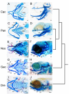
Figure 1
Figure 1
Fish Specimens. Cleared and stained skeletal preparations of the Antarctic silverfish (P. antarcticum), the blackfin icefish (C. aceratus), the yellowbelly rockcod (N. coriiceps), the threespine stickleback (G. aculeatus), and the zebrafish (D. rerio) at comparable stages of development. Bone is stained red with Alizarin red and cartilage is stained blue with Alcian blue. (A, B) C. aceratus, mid-larvae; (C, D)P. antarcticum, mid-larvae; (E, F) N. coriiceps, mid-larvae; (G, H) G. aculeatus, 10 dpf; (I, J) D. rerio, 6 dpf. Both ventral flat mounted (A, C, E, G, I) and lateral (B, D, F, H, J) views are shown. Note the lack of bony elements in pelagic notothenioid species compared to the benthic notothenioid and outgroups. The tree to the right of the images indicates evolutionary relationships. Abbreviations: bsr, branchiostegal rays; cbs, ceratobranchal cartilages; cb5, 5th ceratobrachial cartilage; ch, ceratohyal; cl, cleithrum; co, corocoid; dnt, dentary; ed, endochondral disk; fr, fin rays; hs, hyosymplectic; me, Meckel's cartilage; mx, maxilla; nc, neurocranium; op, opercle; ov, otic vesicle; pmx, premaxilla; pq, palatoquadrate; pt, pterygoid process of the palatoquadrate; te, teeth. Scale bars, 100 μm.
Figure 2
Figure 2
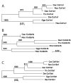
Neighbor-joining tree for col1a1 (A), col2a1b (B) and col10a1 (C). Numbers on internodes are the frequency of each branch in 1,000 bootstrap replicates. Cac = C. aceratus (col1a1, FJ932592; col2a1b, FJ932595; col10a1, FJ932598), Nco = N. coriiceps (col1a1, FJ932593; col2a1b, FJ932596; col10a1, FJ932599), Gac = G. aculeatus (col1a1, FJ932594; col2a1b, FJ932597; col10a1, FJ932600), Tru = Takifugu rubripes (col1a1, N00041; col2a1b, N000077; col10a1, N000010), Dre = Danio rerio (col1a1, NP_954684; col2a1b, BC059180, col10a1, NP_001077296), Gga = Gallus gallus (col1a1, P02457; col2a1b, NP_989757; col10a1, P08125), Hsa = Homo sapiens (col1a1, NP_000079; col2a1b, NP_149162; col10a1, NP_000484).
Figure 3
Figure 3
Whole-mount in situ hybridization for collagen gene expression in the pharyngeal skeleton. Lateral views are shown of embryos for P. antarcticum, C. aceratus, G. aculeatus, and D. rerio at comparable stages of development. (A, E, I) P. antarcticum, mid-larvae; (B, F, J) C. aceratus, mid-larvae; (C, G, K)G. aculeatus, 11 dpf; and (D, H, L) D. rerio, 5 dpf. Notothenioids retain strong col2a1 expression in the pharyngeal skeleton (A, B) well into larval development compared to both G. aculeatus (C) and D. rerio (D). Conversely, col10a1 (E, F) and col1a1 (I, J) expression is relatively weak or absent in the notothenioids. The tree represents the evolutionary relationship among species. Abbreviations: ch, ceratohyal; cl, cleithrum; dnt, dentary; ep, ethmoid plate; ect, ectopterygoid; me, Meckel's cartilage; mx, maxilla; op, opercle; pmx, premaxilla; te, teeth. Scale bars, 100 μm.
Figure 4
Figure 4
Ceratohyal collagen gene expression in P. antarcticum, C. aceratus, G. aculeatus, and D. rerio detected by in situ hybridization on cryosections. (A-C) P. antarcticum, mid-larvae; (D-F) C. aceratus, mid-larvae; (G-I) G. aculeatus, 11 dpf; (J-L) D. rerio, 6 dpf. Sections through the ceratohyal cartilage (ch) are shown in the ventral view. Notothenioid larvae show ubiquitous expression of col2a1 throughout the ch (A, D), whereas col2a1 expression is restricted to the distal ends of the cartilage in both G. aculeatus (G) and D. rerio (J). The col10a1 gene is expressed in hypertrophic chondrocytes in the ch in G. aculeatus (H) and D. rerio (K), whereas its expression is absent from the ch in P. antarcticum (B) and C. aceratus (E). The col1a1 gene is weakly expressed in the periosteum around the ch of both P. antarcticum (C) and C. aceratus (F) relative to the strong expression observed in G. aculeatus (I)and D. rerio (L). The tree represents the evolutionary relationship among species. Scale bars, 100 μm.
Figure 5
Figure 5
Expression of collagen genes in the pectoral fins of P. antarcticum, C. aceratus, G. aculeatus, and D. rerio embryos, detected by whole-mount in situ hybridization. (A-C) P. antarcticum, mid-larvae; (D-F) C. aceratus, mid-larvae; (G-I) G. aculeatus, 7 dpf; (J-L)D. rerio, 3 dpf. Images are of flat-mounted (A-C, G-L) or vibratome-sectioned (D-F) pectoral fins. Expression of col2a1 is observed in the scapulocoracoid (co) of all species (A, D, G, J). Likewise expression of col10a1 (B, E, H, K) and col1a1 (C, F, I, L) is observed in the cleithrum (cl), and col1a1 is expressed in the fin fold (ff) of all species. C. aceratus exhibits a unique pattern of col10a1 expression in the endochondral disk (ed) (E). Thus, gene expression during fin development appears to be largely conserved among these species. The tree represents the evolutionary relationship among species. Scale bars, 100 μm.
Figure 6
Figure 6
Schematic illustration of notothenioid craniofacial evolution via heterochrony. Panel A summarizes results for the relative patterns of each collagen gene expression during notothenioid larval skeletogenesis. Expression in G. aculeatus and D. rerio follows a typical vertebrate pattern, with col2a1 expressed first in differentiating chondrocytes, followed by a down regulation of col2a1 and subsequent up regulation of the bone markers, col10a1 and col1a1 (A, top panel). The notothenioid pattern is quite different, with sustained high levels of col2a1 expression throughout later periods of larval development, expression of col10a1 limited to a small subset of elements, and weak col1a1 expression throughout the pharyngeal skeleton (A, bottom panel). The x-axis denotes developmental time starting just before chondrocyte differentiation, and the y-axis represents relative (not quantitative) levels of collagen expression. Panels B and C depict a model for anterior-posterior and dorsal-ventral patterning of the pharyngeal skeleton. Compared to stickleback and zebrafish, the developmental program in notothenioid species is shifted toward the ventral components (darkly shaded) of the anterior-most craniofacial elements (purple and blue). This shift results in the development of rostral-caudally expanded Meckel's (purple) and ceratohyal (blue) cartilages, and reduced posterior cartilage elements (i.e., fifth ceratobranchial cartilage, yellow).

Similar articles

Cited by

References

    1. Eastman JT. The nature of the diversity of Antarctic fishes. Polar Biol. 2005;28:93–107. doi: 10.1007/s00300-004-0667-4. - DOI
    1. DeConto RM, Pollard D. Rapid Cenozoic glaciation of Antarctica induced by declining atmospheric CO2. Nature. 2003;421:245–249. doi: 10.1038/nature01290. - DOI - PubMed
    1. DeWitt HH. In: Antarctic Map Folio Series. Bushnell VC, editor. Vol. 15. New York: American Geographical Society; 1971. Coastal and deep-water benthic fishes of the Antarctic; pp. 1–10.
    1. Eastman JT. Antarctic Fish Biology: Evolution in a Unique Environment. San Diego: Academic Press; 1993.
    1. Eastman JT, Clarke A. A comparison of adaptive radiations of Antarctic fish with those of non-Antarctic fish. Milano: Springer-Verlag; 1998.

Publication types

LinkOut - more resources