. 2010;12(1):R2.
doi: 10.1186/ar2900. Epub 2010 Jan 6.

A Rac1 inhibitory peptide suppresses antibody production and paw swelling in the murine collagen-induced arthritis model of rheumatoid arthritis

Affiliations

A Rac1 inhibitory peptide suppresses antibody production and paw swelling in the murine collagen-induced arthritis model of rheumatoid arthritis

Joana Rf Abreu et al. Arthritis Res Ther. 2010.

Abstract

Introduction: The Rho family GTPase Rac1 regulates cytoskeletal rearrangements crucial for the recruitment, extravasation and activation of leukocytes at sites of inflammation. Rac1 signaling also promotes the activation and survival of lymphocytes and osteoclasts. Therefore, we assessed the ability of a cell-permeable Rac1 carboxy-terminal inhibitory peptide to modulate disease in mice with collagen-induced arthritis (CIA).

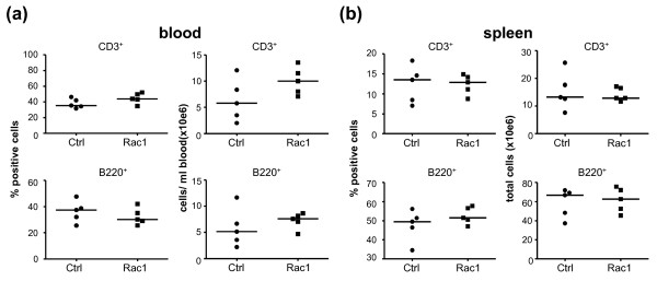
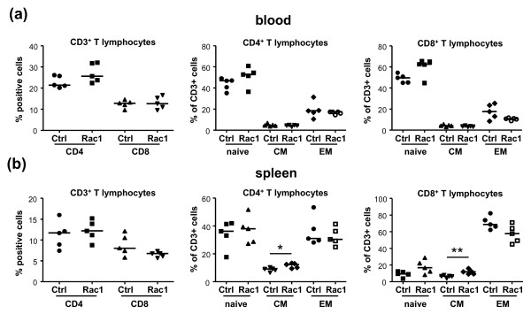
Methods: CIA was induced in DBA/1 mice, and in either early or chronic disease, mice were treated three times per week by intraperitoneal injection with control peptide or Rac1 inhibitory peptide. Effects on disease progression were assessed by measurement of paw swelling. Inflammation and joint destruction were examined by histology and radiology. Serum levels of anti-collagen type II antibodies were measured by enzyme-linked immunosorbent assay. T-cell phenotypes and activation were assessed by fluorescence-activated cell sorting analysis. Results were analyzed using Mann-Whitney U and unpaired Student t tests.

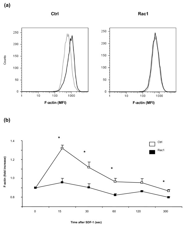
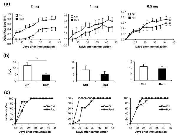
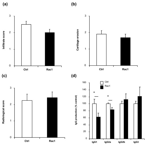
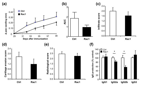
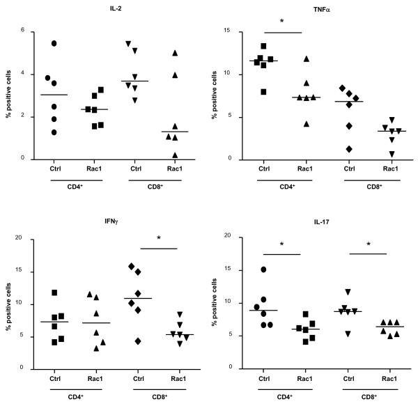
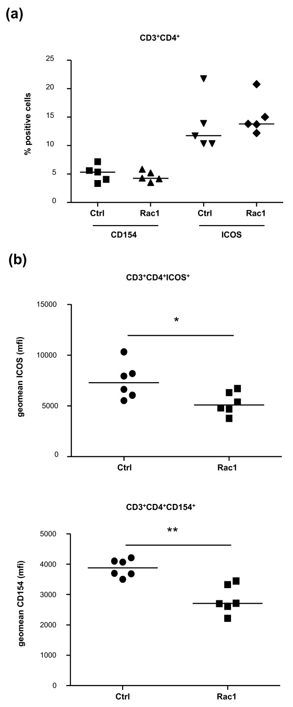
Results: Treatment of mice with Rac1 inhibitory peptide resulted in a decrease in paw swelling in early disease and to a lesser extent in more chronic arthritis. Of interest, while joint destruction was unaffected by Rac1 inhibitory peptide, anti-collagen type II antibody production was significantly diminished in treated mice, in both early and chronic arthritis. Ex vivo, Rac1 inhibitory peptide suppressed T-cell receptor/CD28-dependent production of tumor necrosis factor alpha, interferon gamma and interleukin-17 by T cells from collagen-primed mice, and reduced induction of ICOS and CD154, T-cell costimulatory proteins important for B-cell help.

Conclusions: The data suggest that targeting of Rac1 with the Rac1 carboxy-terminal inhibitory peptide may suppress T-cell activation and autoantibody production in autoimmune disease. Whether this could translate into clinically meaningful improvement remains to be shown.


Figures

Figure 1
Figure 1
The Rac1 carboxy-terminal peptide efficiently blocks actin polymerization in murine cells. (a) Representative histograms of F-actin staining in T cells isolated from mice spleens that were exposed for 15 minutes to 200 μg/mL of Ctrl or Rac1 peptide followed by stromal cell-derived factor 1 (SDF-1) stimulation. (b) Fold increase in F-actin. Data are depicted as mean fold increase (MFI) of F-actin ± standard error of the mean (n = 3). *P < 0.05. Ctrl, control.
Figure 2
Figure 2
Reduced paw swelling, but no effect on disease incidence, after treatment with 2 mg of Rac1 carboxy-terminal peptide. Mice were treated with 2 mg, 1 mg, or 0.5 mg of Ctrl (white square) or Rac1 (black square) peptide at the indicated time points. Paw swelling and inflammation of the four limbs were determined for each mouse. (a) Delta hind paw ankle joint swelling was calculated by subtracting the paw diameter on the day of initiation of treatment from the measured diameter. Values are presented as mean ± standard error of the mean (SEM) (n = 8). (b) Area under the curve (AUC) was calculated for the delta paw swelling in each mouse. Values are presented as mean AUC ± SEM. *P < 0.05. (c) Cumulative incidence of arthritis in mice for each dosage of Ctrl and Rac1 peptide was calculated. Ctrl, control.
Figure 3
Figure 3
Effects of treatment with the Rac1 carboxy-terminal peptide at onset of disease in collagen-induced arthritis. Mice were treated with 2 mg of Ctrl (white bars) or 2 mg of Rac1 (black bars) peptide starting at day 20. After sacrifice, sections from mice paws (n = 8 per group) were stained with hematoxylin and eosin and assessed for (a) cellular infiltration and (b) cartilage erosion. (c) X-rays of hind paws were analyzed for bone damage. (d) Sera from mice that started treatment at day 20 (n = 8) with Ctrl (white bars) or Rac1 (black bars) peptide were collected, and the levels of specific anti-collagen IgG were detected. IgG levels in the sera of Ctrl-treated mice were set to 100%, and the levels obtained in the sera of Rac1-treated mice were then calculated relative to Ctrl. Represented IgG values were calculated within linear regions of the serum dilution curve. All values are presented as mean ± standard error of the mean. *P ≤ 0.05. Ctrl, control.
Figure 4
Figure 4
Effect of Rac1 carboxy-terminal peptide treatment of mice with chronic arthritis. (a) Delta hind paw ankle joint swelling of mice that started treatment at day 29 with 4 mg of Ctrl or Rac1 peptide (three times weekly). Values are representative of two independent experiments (n = 16 per group). (b) Area under the curve (AUC) calculated as in Figure 2b. Paraffin-embedded sections of the hind paws were stained with hematoxylin and eosin and analyzed for (c) synovial inflammation and (d) cartilage destruction. (e) X-rays were analyzed for bone damage (n = 16 per group). (f) Sera from mice that started treatment at day 29 (n = 7) with Ctrl (white bars) or Rac1 (black bars) peptide were collected, and the levels of specific anti-collagen IgG were detected. IgG levels in the sera of Ctrl-treated mice were set to 100%, and the levels obtained in the sera of Rac1 peptide-treated mice were then calculated relative to Ctrl. Represented values were calculated within linear regions of the serum dilution curve. All values are expressed as mean ± standard error of the mean. *P ≤ 0.05. Ctrl, control.
Figure 5
Figure 5
Effect of Rac1 carboxy-terminal peptide treatment of mice on lymphocyte numbers and percentages in blood and spleen. Mice treated with Ctrl or Rac1 peptide (2 mg, n = 5 per group) were sacrificed on day 28 following arthritis induction, and CD3+ T cells and B220+ B cells were identified by fluorescence-activated cell sorting analysis in (a) blood and (b) spleen. Numbers of CD3+ T cells (upper panels) and B220+ cells (lower panels) were calculated as percentage positive cells (left panels) or total number of cells (right panels) per milliliter of blood or per spleen. Data points within each column represent individual mice, and the bar indicates the median value. Ctrl, control.
Figure 6
Figure 6
T-cell phenotyping in arthritic mice treated with Rac1 carboxy-terminal peptide. Blood (a) and spleens (b) were obtained from arthritic mice treated with Ctrl or Rac1 peptide (2 mg, n = 5 per group), and cells were stained with antibodies against CD3, CD4, CD8, CD44, and CD62L. The percentages of CD3+ T cells expressing CD4 and CD8 (left panels) and naïve (CD44-CD62L+), central memory (CM) (CD44+CD62L+), and effector/memory (EM) (CD44+CD62L-) CD3+CD4+ (middle panels) and CD3+CD8+ T cells were calculated. Data points within each column represent individual mice, and the bar indicates the median value. *P ≤ 0.05, *P ≤ 0.01. Ctrl, control.
Figure 7
Figure 7
Effect of Rac1 carboxy-terminal peptide treatment on T-cell cytokine production. Splenocytes obtained from mice (n = 6) on day 19 following priming with bovine collagen type II in complete Freund's adjuvant were preincubated for 15 minutes with Ctrl or Rac1 peptide (200 μg/mL) and stimulated for 24 hours with anti-CD3 and anti-CD28 antibodies. Brefeldin A was included for the last 4 hours of stimulation, and cells were stained for CD3, CD4, CD8, interleukin (IL)-2, tumor necrosis factor-alpha (TNFα), interferon-gamma (IFNγ), and IL-17. The percentages of CD3+CD4+ and CD3+CD8+ T cells expressing each cytokine were determined by fluorescence-activated cell sorting analysis. Data points within each column represent values obtained from splenocytes of individual mice, and the bar indicates the median value. *P ≤ 0.05. Ctrl, control.
Figure 8
Figure 8
Effect of Rac1 carboxy-terminal peptide treatment on T-cell costimulatory protein expression. Splenocytes from mice primed with bovine collagen type II in complete Freund's adjuvant analyzed in Figure 7 (n = 6) were examined for ICOS and CD154 expression by fluorescence-activated cell sorting analysis following 24-hour stimulation with anti-CD3 and anti-CD28 antibodies in the presence of Ctrl or Rac1 peptide (200 μg/mL). The percentages of CD3+CD4+ T cells expressing CD154 and ICOS (a) and the mean geometric (geomean) fluorescence intensity (mfi) of CD154 and ICOS expression on these cells (b) were calculated following fluorescence-activated cell sorting analysis. Data points within each column represent values obtained from splenocytes of individual mice, and the bar indicates the median value. *P ≤ 0.05; **P < 0.01. Ctrl, control.

Comment in

References

    1. Vergunst CE, Sande MG van de, Lebre MC, Tak PP. The role of chemokines in rheumatoid arthritis and osteoarthritis. Scand J Rheumatol. 2005;34:415–425. doi: 10.1080/03009740500439159. - DOI - PubMed
    1. McInnes IB, Schett G. Cytokines in the pathogenesis of rheumatoid arthritis. Nat Rev Immunol. 2007;7:429–442. doi: 10.1038/nri2094. - DOI - PubMed
    1. Jaffe AB, Hall A. Rho GTPases: biochemistry and biology. Annu Rev Cell Dev Biol. 2005;21:247–269. doi: 10.1146/annurev.cellbio.21.020604.150721. - DOI - PubMed
    1. Moll J, Sansig G, Fattori E, Putten H van der. The murine rac1 gene: cDNA cloning, tissue distribution and regulated expression of rac1 mRNA by disassembly of actin microfilaments. Oncogene. 1991;6:863–866. - PubMed
    1. Shirsat NV, Pignolo RJ, Kreider BL, Rovera G. A member of the ras gene superfamily is expressed specifically in T, B and myeloid hemopoietic cells. Oncogene. 1990;5:769–772. - PubMed

Publication types

MeSH terms