

Reduction of CPR artifacts in the ventricular fibrillation ECG by coherent line removal
- PMID: 20053282
- PMCID: PMC2820034
- DOI: 10.1186/1475-925X-9-2
Reduction of CPR artifacts in the ventricular fibrillation ECG by coherent line removal
Abstract
Background: Interruption of cardiopulmonary resuscitation (CPR) impairs the perfusion of the fibrillating heart, worsening the chance for successful defibrillation. Therefore ECG-analysis during ongoing chest compression could provide a considerable progress in comparison with standard analysis techniques working only during "hands-off" intervals.
Methods: For the reduction of CPR-related artifacts in ventricular fibrillation ECG we use a localized version of the coherent line removal algorithm developed by Sintes and Schutz. This method can be used for removal of periodic signals with sufficiently coupled harmonics, and can be adapted to specific situations by optimal choice of its parameters (e.g., the number of harmonics considered for analysis and reconstruction). Our testing was done with 14 different human ventricular fibrillation (VF) ECGs, whose fibrillation band lies in a frequency range of [1 Hz, 5 Hz]. The VF-ECGs were mixed with 12 different ECG-CPR-artifacts recorded in an animal experiment during asystole. The length of each of the ECG-data was chosen to be 20 sec, and testing was done for all 168 = 14 x 12 pairs of data. VF-to-CPR ratio was chosen as -20 dB, -15 dB, -10 dB, -5 dB, 0 dB, 5 dB and 10 dB. Here -20 dB corresponds to the highest level of CPR-artifacts.
Results: For non-optimized coherent line removal based on signals with a VF-to-CPR ratio of -20 dB, -15 dB, -10 dB, -5 dB and 0 dB, the signal-to-noise gains (SNR-gains) were 9.3 +/- 2.4 dB, 9.4 +/- 2.4 dB, 9.5 +/- 2.5 dB, 9.3 +/- 2.5 dB and 8.0 +/- 2.7 (mean +/- std, n = 168), respectively. Characteristically, an original VF-to-CPR ratio of -10 dB, corresponds to a variance ratio var(VF):var(CPR) = 1:10. An improvement by 9.5 dB results in a restored VF-to-CPR ratio of -0.5 dB, corresponding to a variance ratio var(VF):var(CPR) = 1:1.1, the variance of the CPR in the signal being reduced by a factor of 8.9.
Discussion: The localized coherent line removal algorithm uses the information of a single ECG channel. In contrast to multi-channel algorithms, no additional information such as thorax impedance, blood pressure, or pressure exerted on the sternum during CPR is required. Predictors of defibrillation success such as mean and median frequency of VF-ECGs containing CPR-artifacts are prone to being governed by the harmonics of the artifacts. Reduction of CPR-artifacts is therefore necessary for determining reliable values for estimators of defibrillation success.
Conclusions: The localized coherent line removal algorithm reduces CPR-artifacts in VF-ECG, but does not eliminate them. Our SNR-improvements are in the same range as offered by multichannel methods of Rheinberger et al., Husoy et al. and Aase et al. The latter two authors dealt with different ventricular rhythms (VF and VT), whereas here we dealt with VF, only. Additional developments are necessary before the algorithm can be tested in real CPR situations.
Figures

 is displayed for an ECG-signal s = s(t) containing CPR-artifacts. The optimal estimator for the CPR-frequency is taken as the frequency which maximizes this function. Here f is an arbitrary frequency and (kf), k = 2,3, ..., M, are its harmonics. The function ŝ = ŝ(f) is the Fourier transform of the ECG-signal s = s(t) and |ŝ(f)|2 is the powerspectrum. In the present example the optimal estimator f0 for the CPR-frequency is f0 = 1.66 Hz.
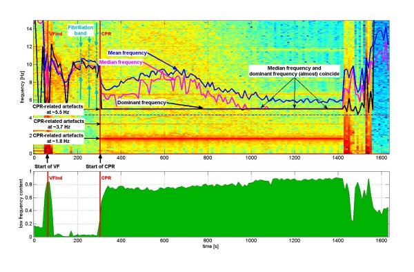


Similar articles
-
A new method to estimate the amplitude spectrum analysis of ventricular fibrillation during cardiopulmonary resuscitation.Resuscitation. 2013 Nov;84(11):1505-11. doi: 10.1016/j.resuscitation.2013.07.004. Epub 2013 Jul 12. Resuscitation. 2013. PMID: 23851191
-
Removal of CPR artifacts from the ventricular fibrillation ECG by adaptive regression on lagged reference signals.IEEE Trans Biomed Eng. 2008 Jan;55(1):130-7. doi: 10.1109/TBME.2007.902235. IEEE Trans Biomed Eng. 2008. PMID: 18232354
-
A new method without reference channels used for ventricular fibrillation detection during cardiopulmonary resuscitation.Australas Phys Eng Sci Med. 2016 Jun;39(2):391-401. doi: 10.1007/s13246-016-0425-2. Epub 2016 Jan 29. Australas Phys Eng Sci Med. 2016. PMID: 26831488
-
[Analysis of ventricular fibrillation signals for the evaluation of defibrillation success in the treatment of ventricular fibrillation].Anasthesiol Intensivmed Notfallmed Schmerzther. 2003 Dec;38(12):787-94. doi: 10.1055/s-2003-45401. Anasthesiol Intensivmed Notfallmed Schmerzther. 2003. PMID: 14666442 Review. German.
-
See through ECG technology during cardiopulmonary resuscitation to analyze rhythm and predict defibrillation outcome.Curr Opin Crit Care. 2016 Jun;22(3):199-205. doi: 10.1097/MCC.0000000000000297. Curr Opin Crit Care. 2016. PMID: 27031917 Review.
Cited by
-
Estimating the amplitude spectrum area of ventricular fibrillation during cardiopulmonary resuscitation using only ECG waveform.Ann Transl Med. 2021 Apr;9(8):619. doi: 10.21037/atm-20-7166. Ann Transl Med. 2021. PMID: 33987317 Free PMC article.
-
Machine learning and feature engineering for predicting pulse presence during chest compressions.R Soc Open Sci. 2021 Nov 10;8(11):210566. doi: 10.1098/rsos.210566. eCollection 2021 Nov. R Soc Open Sci. 2021. PMID: 34804564 Free PMC article.
-
Rhythm analysis during cardiopulmonary resuscitation: past, present, and future.Biomed Res Int. 2014;2014:386010. doi: 10.1155/2014/386010. Epub 2014 Jan 9. Biomed Res Int. 2014. PMID: 24527445 Free PMC article. Review.
-
Automated Condition-Based Suppression of the CPR Artifact in ECG Data to Make a Reliable Shock Decision for AEDs during CPR.Sensors (Basel). 2021 Dec 8;21(24):8210. doi: 10.3390/s21248210. Sensors (Basel). 2021. PMID: 34960308 Free PMC article.
-
Deep Learning Strategy for Sliding ECG Analysis during Cardiopulmonary Resuscitation: Influence of the Hands-Off Time on Accuracy.Sensors (Basel). 2023 May 5;23(9):4500. doi: 10.3390/s23094500. Sensors (Basel). 2023. PMID: 37177703 Free PMC article.
References
Publication types
MeSH terms
LinkOut - more resources
Full Text Sources
Medical
Research Materials
Miscellaneous
