Review
. 2010 May;48(5):851-8.
doi: 10.1016/j.yjmcc.2009.12.017. Epub 2010 Jan 4.

Myofilament length dependent activation

Affiliations
Review

Myofilament length dependent activation

Pieter P de Tombe et al. J Mol Cell Cardiol. 2010 May.

Abstract

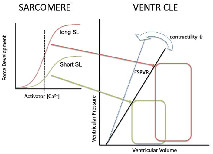
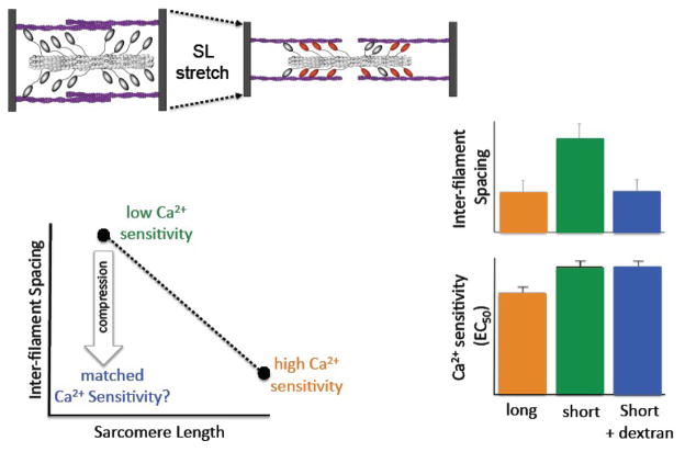
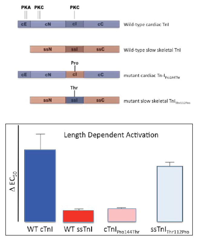
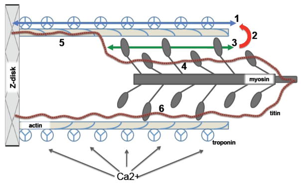
The Frank-Starling law of the heart describes the interrelationship between end-diastolic volume and cardiac ejection volume, a regulatory system that operates on a beat-to-beat basis. The main cellular mechanism that underlies this phenomenon is an increase in the responsiveness of cardiac myofilaments to activating Ca(2+) ions at a longer sarcomere length, commonly referred to as myofilament length-dependent activation. This review focuses on what molecular mechanisms may underlie myofilament length dependency. Specifically, the roles of inter-filament spacing, thick and thin filament based regulation, as well as sarcomeric regulatory proteins are discussed. Although the "Frank-Starling law of the heart" constitutes a fundamental cardiac property that has been appreciated for well over a century, it is still not known in muscle how the contractile apparatus transduces the information concerning sarcomere length to modulate ventricular pressure development.


Figures

Figure 1
Figure 1. The Frank-Starling mechanism and myofilament length dependent activation
The Frank-Starling Law of the Heart describes a fundamental property of the heart (figure on the right). That is, for a given contractile state there is a unique relationship between end-systolic pressure reached in the heart and end-systolic pressure (ESPVR); increased contractility results in an increased slope of the ESPVR (cf. blue arrow). Increased ventricular filling (pre-load; red PV loop) leads to an increase in ventricular pressure development at end-systole which allows for i) increased stroke volume for a given systolic pressure (after-load) and ii) sustained stroke volume at elevated systolic pressure. The Frank-Starling mechanism has, as its basis, a modulation of myofilament Ca2+ sensitivity upon a change in sarcomere length as illustrated in the left graphs. Myofilament force development is the result of activation by Ca2+ ions. The relationship between force development and activator [Ca2+] is shifted up and to the left at longer sarcomere length (short SL, green; long SL, red). For a given contractile state (and, thus, cytosolic [Ca2+]; dashed vertical line), more myofilament force is developed at the longer SL (red) leading to a higher ventricular pressure at higher end-systolic volume (red PV loop). Thus, the Frank-Starling Law of the heart is a direct consequence of the myofilament length dependent activation properties of the cardiac sarcomere.
Figure 2
Figure 2. Schematic of the sarcomere and putative mechanisms underlying myofilament length dependent activation
Schematic diagram depicting the striated muscle sarcomere and some of the possible molecular mechanisms underlying myofilament length dependent activation (indicated by bold numbers). Changes in sarcomere length (SL) occur by means relative sliding between the thin filament (slender tope colored bars) and thick filament (dark grey bars). Thin filaments contains actin, tropomyosin (blue coil) and the troponin complex (composed of TnT, TnC, and TnI; three segmented circles). The globular portion of myosin interacts with actin to generate a mechanical force that is transmitted via the thin filament to the Z-disk and from there to the extra-cellular matrix. Ca2+ binding to troponin activates the thin filament to initiate contraction. Titin is a large molecular weight filament protein that spans between the Z-disk and the M-band of the thick filament. Myofilament length dependent activation could be the result of: i) modulation of pathways involving thin filament activation by troponin, either by directly modulating troponin-troponin cooperative interaction (1) or via a titin-actin interaction modulating the level of thin filament activation, either directly or by altered troponin Ca2+ binding affinity (5). ii) alterations in the cooperative interaction between force generating cross-bridges because the number of attached cross-bridges varies with SL (3); in addition, attached cross-bridges may affect thin filament activation and Ca2+ binding affinity (red arrow; 2), causing an indirect strong binding cross-bridges-thin filament activation feedback modulation that depends on SL. iii) titin interaction with myosin in the A-band may alter the structure of the globular domain of myosin heads that are interacting with actin in a length dependent manner (6). iv) finally, titin interaction with myosin in the pre-force state of the cross-bridge cycle may affect the structure and/or distribution of these weakly bound cross-bridges so as to affect the number of cross-bridges entering the force-generating strongly bound state in a length dependent manner (4).
Figure 3
Figure 3. The inter-filament spacing hypothesis
Modulation of myofilament inter-filament spacing has been proposed as a unifying mechanism for myofilament length dependent activation. Stretch of the sarcomere (top left) is expected to result in a closer approximation of the thick and filaments within the sarcomere (dashed arrows) resulting in a more favorable disposition of myosin heads to interact with actin (illustrated by red myosin heads on the right). Direct measurement[59, 68] of inter-filament spacing by x-ray diffraction has confirmed an inverse relationship between inter-filament spacing and sarcomere length (bottom left). A short sarcomere length is associated with a large inter-filament spacing and low myofilament Ca2+ sensitivity (green), while the opposite is true for a long sarcomere length (orange). Compression of the myofilament lattice (e.g. by dextran) at a short sarcomere length is expected, within the framework of the inter-filament spacing hypothesis, to lead to matched myofilament Ca2+ sensitivity[56, 74] even though sarcomere length has not changed (cf. blue arrow). The bar graphs (lower right) show the results of such an experiment: inter-filament spacing, as measured by x-ray diffraction, was varied by either a change in sarcomere length (long SL, orange; short SL, green) or by compression with 1% dextran (blue). Even though compression resulted in an inter-filament spacing that was matched to that attained at the long sarcomere length (top bars), myofilament Ca2+ sensitivity (as indexed by EC50) was not affected by the osmotic compression (bottom bars). These data, and other data discussed in the text, suggest that inter-filament spacing may not be primary mechanism that underlies myofilament length dependent activation (modified from [71]).
Figure 4
Figure 4. The inhibitory region of cardiac troponin-I plays a pivotal role in myofilament length dependent activation
The adult heart expresses a unique isoform of troponin-I (wild-type cardiac TnI, top diagram; shaded in dark blue) that contains a unique N-terminus 33 amino acid extension (cE). In addition, cardiac TnI contains both PKA and PKC phosphorylation target sites that are lacking in slow skeletal TnI (wild-type slow skeletal TnI; shaded in red). The inhibitory region differs between cardiac TnI and slow skeletal TnI by a single amino acid residue: a threonine at position 144 in cardiac TnI, and a proline at position 112 in slow skeletal TnI. Exchange for recombinant wild-type cardiac troponin containing cardiac TnI into isolated chemically permeabilized myocardium retained the robust myofilament length dependency that is characteristic of this muscle type (cf. bar graphs; DEC50, index of length dependency; WT cTnI; dark blue bar), while exchange for cardiac troponin containing slow skeletal TnI virtually abolished length dependency (WT ssTnI; dark red bar). Exchange for troponin containing a mutant cardiac TnI in which threonine 144 was replaced by a proline (as is found in ssTnI) also virtually eliminated length dependency (cTnIPro144Thr; pink bar), while introduction of a threonine at the equivalent position in slow skeletal TnI markedly enhanced length dependency (ssTnIThr112Pro). These data suggest that presence of a threonine at position 144 is both necessary and sufficient to impart length dependent activation properties upon the cardiac sarcomere (modified from [67]).

Comment in

References

    1. Sagawa K, Maughan L, Suga H, Sunagawa K. Cardiac contraction and the pressure-volume relationship. New York, Oxford: Oxford University Press; 1988.
    1. Pacher P, Nagayama T, Mukhopadhyay P, Batkai S, Kass DA. Measurement of cardiac function using pressure-volume conductance catheter technique in mice and rats. Nat Protoc. 2008;3(9):1422–34. - PMC - PubMed
    1. de Tombe PP. Cardiac myofilaments: mechanics and regulation. J Biomech. 2003 May;36(5):721–30. - PubMed
    1. Solaro RJ. Integration of myofilament response to Ca2+ with cardiac pump regulation and pump dynamics. Am J Physiol. 1999 Dec;277(6 Pt 2):S155–63. - PubMed
    1. ter Keurs HEDJ, Tyberg JV. Mechanics of the Circulation. Dordrecht: Martinus Nijhoff; 1986.

Publication types