. 2010 Jul;80(1):75-88.
doi: 10.1016/j.mvr.2009.12.010. Epub 2010 Jan 4.

Quantitative distribution and colocalization of non-muscle myosin light chain kinase isoforms and cortactin in human lung endothelium

Affiliations

Quantitative distribution and colocalization of non-muscle myosin light chain kinase isoforms and cortactin in human lung endothelium

Mary Brown et al. Microvasc Res. 2010 Jul.

Abstract

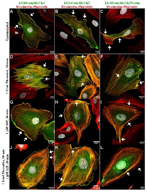
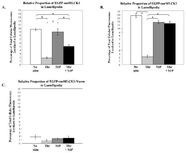
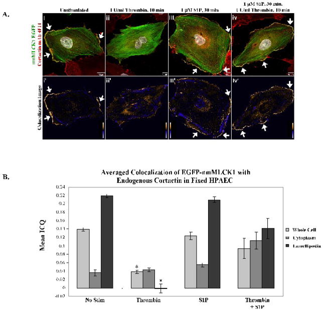
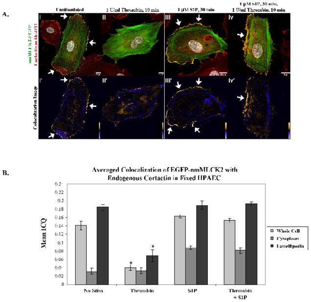
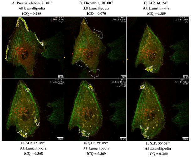
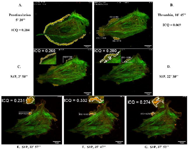
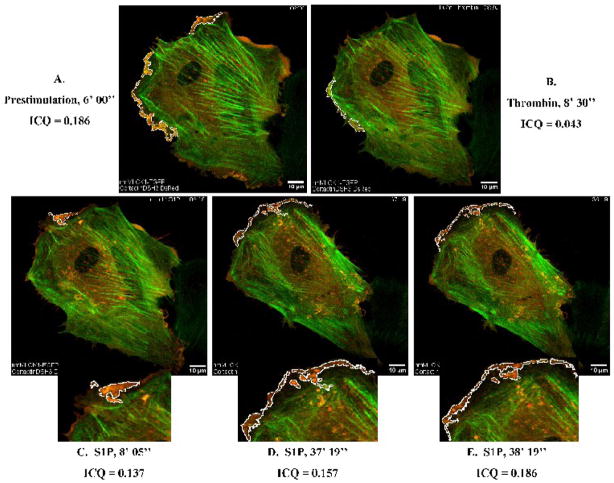
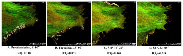
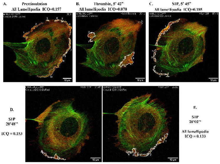
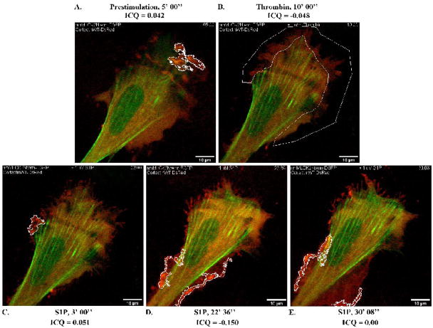
Vascular barrier regulation is intimately linked to alterations in the distribution and configuration of the endothelial cell (EC) cytoskeleton in response to angiogenic and edemagenic agonists. Critical actin cytoskeletal rearrangement includes spatially directed increases in myosin light chain (MLC) phosphorylation, catalyzed by Ca(2+)/calmodulin-dependent non-muscle myosin light chain kinase variants (nmMLCK1- and -2), as well as association of nmMLCK with the actin-binding protein, cortactin. As these associations have proven difficult to quantify in a spatially specific manner, we now describe the utility of intensity correlation image analysis and the intensity correlation quotient (ICQ) to quantify colocalization in fixed and live cell imaging assays in human pulmonary artery EC. From baseline ICQ values averaging 0.216 reflecting colocalization of cortactin-DsRed with EGFP-nmMLCK fusion proteins in resting EC, thrombin-induced EC contraction significantly reduced cortactin-DsRed-EGFP-nmMLCK colocalization (nmMLCK1: ICQ=0.118; nmMLCK2: ICQ=0.091) whereas the potent EC barrier-protective agonist, sphingosine 1-phosphate (S1P), significantly increased nmMLCK-cortactin colocalization within lamellipodia (nmMLCK1: ICQ=0.275; nmMLCK2: ICQ=0.334). Over-expression of a cortactin-DsRed mutant fusion protein lacking the SH3 domain, known to be essential for cortactin-nmMLCK association, reduced baseline and S1P-mediated live cell colocalization with each nmMLCK variant (nmMLCK1: ICQ=0.160; nmMLCK2: ICQ=0.157). Similarly, expression of a truncated EGFP-nmMLCK2 mutant lacking cortactin- and actin-binding domains, markedly reduced basal localization in lamellipodia and abolished colocalization with cortactin-DsRed in lamellipodia after S1P (ICQ=-0.148). These data provide insights into the molecular basis for vascular barrier-regulatory cytoskeletal responses and support the utility of sophisticated imaging analyses and methodological assessment to quantify the critical nmMLCK and cortactin interaction during vascular barrier regulation.


Figures

Figure 1
Figure 1. Distribution of nmMLCK variants and mutant fusion proteins in human lung endothelium
EGFP conjugates of the nmMLCK1 and nmMLCK2 variants, as well as the 496-aa N-terminal fragment of nmMLCK2 were ectopically expressed in human lung EC and incubated with either vehicle (Panels A, B, C), thrombin for 10 min (Panels D, E, F), S1P for 30 min (Panels G, H, I), or thrombin for 10 min and followed by S1P for 30 min (Panels J, K, L). EC were then fixed, immunostained, and imaged as described in Methods section. In unstimulated cells (Panels A, B, C), EGFP-nmMLCK1 and -2 lie along thin fibers transversing the cell as well as strongly localized within lamellipodia (arrows). The N-terminal fragment of nmMLCK2 localizes to broad actin fibers but fails to localize within lamellipodia (Panel C). Thrombin challenge causes EC retraction and disappearance of lamellipodia and redistribution of both nmMLCK variants as well as nmMLCK2Nterm to thick actin stress fibers (arrows, Panels D, E, F). S1P results in translocation of some nmMLCK to spatially distinct actin cortical bands (Panels G, H, J, K) along with ruffling lamellipodia (arrows). The N-terminal fragment of nmMLCK2 is largely absent from lamellipodia (Panels I and L).
Figure 2
Figure 2. Relative proportion of nmMLCK isoforms in human lung endothelial lamellipodia
Human lung EC were treated as described in Figure 1, and the percentage of total spatially–specific cellular distribution of nmMLCK isoforms determined (see Methods) after vehicle, thrombin for 10 min, S1P for 30 min, or thrombin for 10 min followed by S1P for 30 min. Panel A: The percentage of total cellular EGFP-nmMLCK1 localized to lamellipodia is indicated for each condition. * p<0.05; N = 4–18 per condition. Panel B: The percentage of total cellular EGFP-nmMLCK2 localized to lamellipodia is indicated for each condition. * p<0.05; N = 7–17 per condition. Panel C: The percentage of total cellular EGFP-nmMLCK2Nterm localized to lamellipodia is indicated for each condition. Less than 2% is localized to lamellipodia in each condition. N = 10–14 per condition.
Figure 3
Figure 3. Colocalization of EGFP-nmMLCK1 and endogenous cortactin in human lung endothelial lamellipodia
Panel A. EC overexpressing EGFP-nmMLCK1 were incubated with either vehicle (i), thrombin for 10 min (ii), S1P for 30 min (iii), or thrombin for 10 min followed by S1P for 30 min (iv) at 37°C, then fixed and immunostained for cortactin (red). Yellow indicates areas of colocalization. The lower panels, i-iv′, depict colocalization of endogenous cortactin with nmMLCK1. Warm colors indicate pixels with positive mean deviation products, where cortactin and nmMLCK1 fluorescence intensities rose in synchrony above the cellular average intensity, thus indicating colocalization. Blue pixels represent regions with negative mean deviation products in which the cortactin and nmMLCK1 fluorescence intensities varied independently of each other. Shown are representative images selected from 4–20 images for each treatment. Panel B: The mean ICQ values for cortactin and EGFP-nmMLCK1 colocalization were calculated as described in Methods for the entire cell, the cytoplasm, and the lamellipodia under each condition [vehicle (No stim), thrombin for 10 min, S1P for 30 min, or thrombin for 10 min followed by S1P for 30 min]. Thrombin induces a statistically significant reduction in peripheral as well as whole-cell colocalization of nmMLCK1 with cortactin when compared to unstimulated (whole cell or lamellipodia, *p <0.001) and S1P-stimulated cells (whole cell or lamellipodia, *p <0.004). N = 4–20 per condition.
Figure 4
Figure 4. Colocalization of EGFP-nmMLCK2 and endogenous cortactin in human lung endothelial lamellipodia
Panel A. EC overexpressing EGFP-nmMLCK2 were incubated with either vehicle (i), thrombin for 10 min (ii), S1P for 30 min (iii), or thrombin for 10 min followed by S1P for 30 min (iv) at 37°C, then fixed and immunostained for cortactin (red). Yellow indicates areas of colocalization. The lower panels, i-iv′, depict colocalization of endogenous cortactin with nmMLCK2 as described for Figure 3 above. Shown are representative images selected from 7–13 images for each treatment. Panel B: The mean ICQ values for cortactin and EGFP-nmMLCK2 colocalization were calculated as described in Methods for the entire cell, the cytoplasm, and the lamellipodia under each condition [vehicle (No stim), thrombin for 10 min, S1P for 30 min, or thrombin for 10 min followed by S1P for 30 min]. Thrombin induces a statistically significant reduction in peripheral as well as whole-cell colocalization of nmMLCK2 with cortactin when compared to unstimulated (whole cell or lamellipodia, *p <0.02) and S1P-stimulated cells (whole cell or lamellipodia, *p <0.01). N = 7–13 per condition.
Figure 5
Figure 5. Dynamic colocalization of actin-GFP and cortactin-DsRed with intensity correlation analysis applied to live-cell imaging
HPAEC were cotransfected with actin-GFP and cortactin-DsRed constructs and then subjected to live cell imaging as described in Methods. The panels represent images from an extended movie (see Supplemental Movie 1) depicting dynamic colocalization of cortactin-DsRed and actin-GFP in a single EC under basal conditions (A), after thrombin stimulation (B), and subsequent S1P stimulation (C–F). Yellow indicates areas of colocalization. ICQ quantitation for actin-cortactin colocalization in outlined lamellipodia is shown for each panel.
Figure 6
Figure 6. Dynamic colocalization of EGFP-nmMLCK1 and cortactin-DsRed in live human lung EC
HPAEC were cotransfected with EGFP-nmMLCK1 and cortactin-DsRed constructs and then subjected to live cell imaging as described in Methods. The panels represent images from an extended movie (see Supplemental Movie 2) depicting dynamic colocalization of cortactin-DsRed and EGFP-nmMLCK1 in a single EC under basal conditions (A), after thrombin stimulation (B), and subsequent S1P stimulation (C–G). Yellow indicates areas of colocalization. ICQ quantitation for nmMLCK1-cortactin colocalization in outlined lamellipodia is shown for each panel.
Figure 7
Figure 7. Reduced colocalization of cortactinΔSH3-DsRed with EGFP-nmMLCK1 in live human lung EC
HPAEC were cotransfected with EGFP-nmMLCK1 and cortactinΔSH3-DsRed constructs and then subjected to live cell imaging as described in Methods. The panels represent images from an extended movie (see Supplemental Movie 3) depicting dynamic colocalization of cortactinΔSH3-DsRed and EGFP-nmMLCK1 in a single EC under basal conditions (A), after thrombin stimulation (B), and subsequent S1P stimulation (C–E). Yellow indicates areas of colocalization. ICQ quantitation for nmMLCK1-cortactinΔSH3 colocalization in outlined lamellipodia is shown for each panel.
Figure 8
Figure 8. Dynamic colocalization of EGFP-nmMLCK2 and cortactin-DsRed in lamellipodia
HPAEC were cotransfected with EGFP-nmMLCK2 and cortactin-DsRed constructs and then subjected to live cell imaging as described in Methods. The panels represent images from an extended movie (see Supplemental Movie 4) depicting dynamic colocalization of cortactin-DsRed and EGFP-nmMLCK2 in a single EC under basal conditions (A), after thrombin stimulation (B), and subsequent S1P stimulation (C–D). Yellow indicates areas of colocalization. ICQ quantitation for nmMLCK2-cortactin colocalization in outlined lamellipodia is shown for each panel.
Figure 9
Figure 9. Reduced colocalization of cortactinΔSH3-DsRed with EGFP-nmMLCK2 in live human lung EC
HPAEC were cotransfected with EGFP-nmMLCK2 and cortactinΔSH3-DsRed constructs and then subjected to live cell imaging as described in Methods. The panels represent images from an extended movie (see Supplemental Movie 5) depicting dynamic colocalization of cortactinΔSH3-DsRed and EGFP-nmMLCK2 in a single EC under basal conditions (A), after thrombin stimulation (B), and subsequent S1P stimulation (C–E). Yellow indicates areas of colocalization. ICQ quantitation for nmMLCK2- cortactinΔSH3 colocalization in outlined lamellipodia is shown for each panel.
Figure 10
Figure 10. Deletion of the actin- and cortactin-binding domains of nmMLCK2 ablates colocalization with cortactin in S1P-stimulated live EC
HPAEC were cotransfected with EGFP-nmMLCK2Nterm and cortactin-DsRed constructs and then subjected to live cell imaging as described in Methods. The panels represent images from an extended movie (see Supplemental Movie 6) depicting dynamic colocalization of cortactin-DsRed and EGFP-nmMLCK2Nterm in a single EC under basal conditions (A), after thrombin stimulation (B), and subsequent S1P stimulation (C–E). Yellow indicates areas of colocalization. ICQ quantitation for nmMLCK2Nterm-cortactin colocalization in outlined lamellipodia is shown for each panel.

Similar articles

Cited by

References

    1. Birukov KG, et al. Differential regulation of alternatively spliced endothelial cell myosin light chain kinase isoforms by p60(Src) J Biol Chem. 2001;276:8567–73. - PubMed
    1. Bolte S, Cordelieres FP. A guided tour into subcellular colocalization analysis in light microscopy. Journal of Microscopy. 2006;224:213–232. - PubMed
    1. Brittain JM, et al. An atypical role for collapsin response mediator protein 2 (CRMP-2) in neurotransmitter release via interaction with presynaptic voltage-gated calcium channels. J Biol Chem. 2009;284:31375–90. - PMC - PubMed
    1. Burkhardt JK, et al. Overexpression of the dynamitin (p50) subunit of the dynactin complex disrupts dynein-dependent maintenance of membrane organelle distribution. Journal of Cell Biology. 1997;139:469–484. - PMC - PubMed
    1. Burkhardt JK, et al. The giant organelles in Beige and Chediak-Higashi Fibroblasts are derived from late endosomes and mature lysosomes. Journal of Experimental Medicine. 1993;178:1845–1856. - PMC - PubMed

Publication types

MeSH terms