

Quantitative assessment of conjunctival microvascular circulation of the human eye
- PMID: 20053367
- PMCID: PMC3253734
- DOI: 10.1016/j.mvr.2009.12.003
Quantitative assessment of conjunctival microvascular circulation of the human eye
Abstract
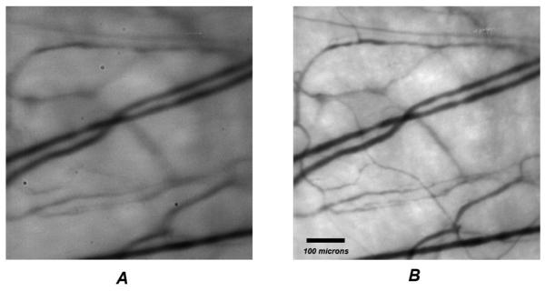
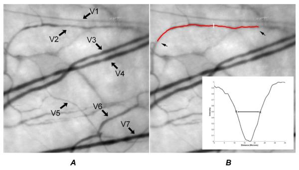
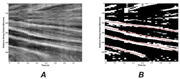
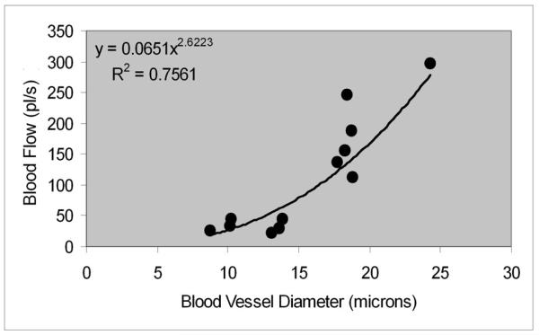
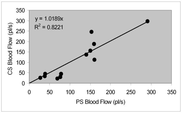
Accessibility to the bulbar conjunctival microvasculature provides a means to assess blood supply to the cerebral cortex and thus optimize therapeutic interventions designed to prevent or reduce the risk of cerebral vascular disease and stroke. The feasibility of a method for quantitative measurements of conjunctiva blood vessel diameter, blood velocity, and flow in the human eye is reported. The method is based on slit lamp biomicroscope digital imaging coupled with a space time image analysis technique. A sequence of conjunctiva microvasculature images was captured at a rate of 50 Hz. The images were analyzed to determine blood vessel diameter, velocity and flow. Blood vessel diameter measurements ranged between 8.7 and 24.3 microns, with a mean value of 15.5 microns. Blood flow rate ranged between 27.3 and 296.9 pl/s, with a mean value of 111.8 pl/s. The relationship between blood flow and vessel diameter was fit with a power law curve (R=0.87). The application of this technique for in vivo quantitative assessment of blood flow dynamics has potential to impact diagnosis and monitoring of various cardiovascular and blood disorders.
Copyright 2009 Elsevier Inc. All rights reserved.
Figures
Similar articles
-
Vessel Sampling and Blood Flow Velocity Distribution With Vessel Diameter for Characterizing the Human Bulbar Conjunctival Microvasculature.Eye Contact Lens. 2016 Mar;42(2):135-40. doi: 10.1097/ICL.0000000000000146. Eye Contact Lens. 2016. PMID: 25839347 Free PMC article.
-
Quantitative assessment of the conjunctival microcirculation using a smartphone and slit-lamp biomicroscope.Microvasc Res. 2019 Nov;126:103907. doi: 10.1016/j.mvr.2019.103907. Epub 2019 Jul 19. Microvasc Res. 2019. PMID: 31330150
-
Measurement variability of the bulbar conjunctival microvasculature in healthy subjects using functional slit lamp biomicroscopy (FSLB).Microvasc Res. 2015 Sep;101:15-9. doi: 10.1016/j.mvr.2015.05.003. Epub 2015 Jun 16. Microvasc Res. 2015. PMID: 26092682 Free PMC article.
-
Conjunctival microcirculation in ocular and systemic microvascular disease.Clin Exp Optom. 2023 Sep;106(7):694-702. doi: 10.1080/08164622.2022.2151872. Epub 2023 Jan 15. Clin Exp Optom. 2023. PMID: 36641840 Review.
-
Progress of Bulbar Conjunctival Microcirculation Alterations in the Diagnosis of Ocular Diseases.Dis Markers. 2022 Aug 28;2022:4046809. doi: 10.1155/2022/4046809. eCollection 2022. Dis Markers. 2022. PMID: 36072898 Free PMC article. Review.
Cited by
-
Development of a perfusable, hierarchical microvasculature-on-a-chip model.Lab Chip. 2023 Oct 10;23(20):4552-4564. doi: 10.1039/d3lc00512g. Lab Chip. 2023. PMID: 37771308 Free PMC article.
-
Retinal vessel diameter assessment in papilledema by semi-automated analysis of SLO images: feasibility and reliability.Invest Ophthalmol Vis Sci. 2014 Apr 3;55(4):2049-54. doi: 10.1167/iovs.13-13621. Invest Ophthalmol Vis Sci. 2014. PMID: 24609623 Free PMC article.
-
Automated Assessment of Hemodynamics in the Conjunctival Microvasculature Network.IEEE Trans Med Imaging. 2016 Feb;35(2):605-11. doi: 10.1109/TMI.2015.2486619. Epub 2015 Oct 6. IEEE Trans Med Imaging. 2016. PMID: 26452274 Free PMC article.
-
Human conjunctival microvasculature assessed with a retinal function imager (RFI).Microvasc Res. 2013 Jan;85:134-7. doi: 10.1016/j.mvr.2012.10.003. Epub 2012 Oct 16. Microvasc Res. 2013. PMID: 23084966 Free PMC article.
-
Evaluation of regional bulbar redness using an image-based objective method.Int J Ophthalmol. 2014 Feb 18;7(1):71-6. doi: 10.3980/j.issn.2222-3959.2014.01.13. eCollection 2014. Int J Ophthalmol. 2014. PMID: 24634867 Free PMC article.
References
-
- Alizade IT. Acoustic dysfunction and microcirculation in patients with diabetes mellitus. Vestn Otorinolaringol. 2007:11–13. - PubMed
-
- Cheung AT, et al. Microvascular abnormalities in sickle cell disease: a computer-assisted intravital microscopy study. Blood. 2002;99:3999–4005. - PubMed
-
- Cheung AT, et al. Correlation of abnormal intracranial vessel velocity, measured by transcranial Doppler ultrasonography, with abnormal conjunctival vessel velocity, measured by computer-assisted intravital microscopy, in sickle cell disease. Blood. 2001;97:3401–3404. - PubMed
-
- Duench S, et al. Assessment of variation in bulbar conjunctival redness, temperature, and blood flow. Optom Vis Sci. 2007;84:511–516. - PubMed
-
- Ellis CG, et al. Application of image analysis for evaluation of red blood cell dynamics in capillaries. Microvasc Res. 1992;44:214–225. - PubMed
Publication types
MeSH terms
Grants and funding
LinkOut - more resources
Full Text Sources
Other Literature Sources
